
What’s Really Happening Inside the Capsid of Norovirus VLPs?
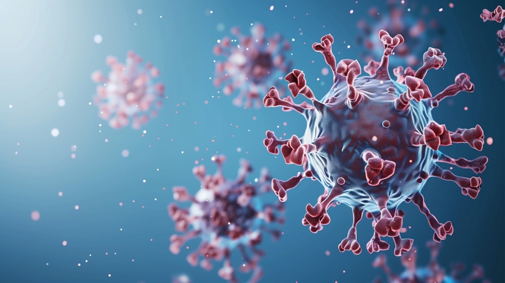
Norovirus virus-like particles (VLPs) are structurally heterogeneous, built from a single capsid protein that self-assembles into multiple geometries, making full characterization by conventional mass spectrometry (MS) extremely difficult.
A recent bioRxiv paper by Dr. Charlotte Uetrecht and colleagues at the Centre for Structural Systems Biology (CSSB) in Hamburg, Germany, in collaboration with Waters scientists, demonstrates how charge detection mass spectrometry (CDMS) is pivotal for uncovering a non-classical capsid assembly invisible to any other method.
In the study, CDMS was used to analyze two human norovirus strains, with cryo-EM and bottom-up proteomics providing orthogonal validation.
The core problem: Heterogeneity defeats conventional MS
Norovirus VLPs can form T=1, T=3, T=4, and other geometries. Combined with N-terminal processing of the VP1 capsid protein, this creates overlapping mass distributions that conventional native MS cannot resolve—coexisting particle populations, including non-icosahedral species, are entirely obscured.
How CDMS solves the problem
CDMS determined the mass and charge of individual ions simultaneously, by passing the need for charge state resolution. The authors compared results obtained from the electrostatic linear ion trip (ELIT)-based Xevo CDMS and the Orbitrap-based Direct Mass Technology (DMT) and reported that:
- Both platforms identified the same major assemblies in GI.1 Norwalk and GII.17 Kawasaki VLPs
- Waters Xevo CDMS produced masses closer to theoretical values, attributed to its dual m/z and charge calibration that accounts for adduct mass
- DMT combined with UniDec deconvolution (UCD) achieved higher apparent mass resolution, at the cost of greater workflow complexity and risk of deconvolution artifacts
CDMS and cryo-EM jointly reveal a Novel GII.17 capsid
Combining CDMS and cryo-EM enabled discovery and structural validation of a novel viral assembly state that would have been difficult to identify with either technique alone.
- CDMS detected three distinct capsid populations in GII.17 Kawasaki: classical T=3 (~10.65 MDa), T=4 (~14.25 MDa), and an unexpected intermediate species (~12.58 MDa).
- Mass analysis estimated ~212 VP1 subunits for the intermediate particle, suggesting a non-icosahedral assembly inconsistent with traditional Caspar-Klug symmetry.
- cryo-EM confirmed the structural identity of this species as a prolate (oval-shaped) capsid and yielded the first images of GII.17 Kawasaki T=3, T=4, and prolate assemblies.
Instrument comparison guides future strategy
Stepwise inlet heating in the Xevo CDMS reduced measured mass toward theoretical values—a practical tool for separating true particle mass from adduct contributions.
Key takeaways:
- Xevo CDMS offers a streamlined workflow with live data visualization and mass accuracy within ±1%
- DMT plus UCD deconvolution reduces peak width by ~50%, but requires careful application to avoid artifacts
- Analyte-matched calibration standards are critical—both platforms showed mass deviations linked to calibrant size mismatch
Read the full paper, “Applying distinct CDMS strategies to observe non-classical virus capsid assembly”, to see how two CDMS platforms together reveal the full complexity of norovirus VLP populations.
Related posts
PROTACs Are Different: What Does That Mean for Liquid Chromatography?
As drug discovery moves beyond traditional small-molecule inhibitors, analytical scientists are being asked to characterize increasingly complex molecules. Proteolysis-targeting chimeras (PROTACs) are one example. PROTACs combine two binding elements with a linker to selectively target proteins for degradation. Their larger size, flexibility, and multiple functional groups can create new analytical considerations compared to more conventional…
Breaking the Megadalton Barrier: Benchtop CDMS Arrives
A recent bioRxiv paper by Dr. Jakub Ujma and colleagues at Waters Corporation, in collaboration with Prof. Martin Jarrold from Indiana University and Megadalton Solutions, describes a major advance—the first commercial benchtop charge detection mass spectrometer (CDMS) based on electrostatic linear ion trap (ELIT) technology. Why does this matter CDMS is uniquely suited for analyzing…

Advancing Oligonucleotide Purification with MaxPeak Premier OBD Prep Columns
Oligonucleotide therapeutics continue to grow in complexity, and so do the purification challenges that come with them. Scientists need reliable, scalable solutions for preparative oligonucleotide purification. MaxPeak Premier Oligo OBD Columns provide a powerful solution for these workflows, now expanded to include 19mm ID formats. These columns reduce non-specific adsorption, improve recovery, and enable consistent…
Popular Topics
ACQUITY QDa (17) bioanalysis (13) biologics (17) biopharma (50) biopharmaceutical (59) biotherapeutics (24) case study (22) chromatography (16) data integrity (28) HPLC (17) informatics (12) LC-MS (35) liquid chromatography (LC) (34) mass detection (16) mass spectrometry (MS) (68) method development (16) particle analysis (21) regulatory compliance (16)