High-Throughput LNP Compositional Analysis Using GTxResolve™ RP 230 Å PH+ Columns: Method Development Considerations
Mateusz Imiolek, Stephan M. Koza
Waters Corporation, United States
Published on June 08, 2026
Abstract
This application note introduces a new GTxResolve Reversed Phase Column, designed for the separation of highly hydrophobic analytes such as lipids, and demonstrates its utility for compositional analysis of lipid nanoparticles (LNPs) while providing practical method development guidance. The columns are packed with wide-pore (230 Å) superficially porous particles engineered to deliver optimal retention for lipid separations through a combination of high surface charge density and a phenyl-hexyl bonded phase. This application note shows that the unique ligand chemistry imparts a positively charged particle surface under acidic conditions, facilitating method development. Particularly for ionizable lipid species, where the electrostatic interactions can be readily modulated by adjusting the mobile phase ionic strength (0–10 mM), providing a convenient and effective optimization parameter. In addition, the phenyl-hexyl ligand enables fine control of selectivity through the incorporation of low levels (~30%) of protic organic modifiers, with MeOH demonstrating a pronounced effect. This study highlights how those insights were applied to optimize the separation of a challenging LNP formulation and provide general guiding principles for lipid class-selective separations. Altogether, this application note demonstrates how the GTxResolve RP 230 Å PH+ Columns design can be leveraged to address complex LNP and lipid separations and support regulatory driven characterization requirements.
Benefits
- Demonstrates how a new wide pore particle design allows for high-throughput lipid and LNP separations
- Highlights the advantages of a positively charged particle surface for optimizing separations of ionizable (cationic) lipids
- Establishes practical method development guidelines based on mobile phase ionic strength and organic modifier selection, emphasizing the key role of MeOH
Introduction
Lipids have become an important component of modern drug delivery systems due to their proven biocompatibility and versatile payload encapsulation capabilities. The landscape of specialized delivery vehicles originated with liposomes but now extends to various nanostructured lipid carriers, (solid) lipid nanoparticles, lipoplexes and non-lamellar lipid nanoparticles developed for delivery of multiple therapeutic modalities, including small molecules, nucleic acids and peptides.1 Such diversity necessitates the use of lipids with diverse physicochemical properties, which can pose specific challenges in routine analytical assays. For example, certain hydrophobic lipids are difficult to elute from generic C18 stationary phases under standard mobile phase conditions.2 Nevertheless, as components of complex drug products, all constituents may need to be comprehensively characterized, both as raw materials and within the final drug product (e.g. stability studies).3 Similarly, the usefulness of lipidomics methods is generally evaluated by their breadth of lipid coverage, robustness, and discriminating power. Addressing these challenges requires the use of fit-for-purpose and well-understood chromatographic tools.
The critical quality attributes (CQAs) of identification and quantification of lipid components and impurities are typically measured using reversed phase chromatography. While it is considered the method of choice due to its capability of resolving intraclass and interclass lipid species,4 it often requires lengthy trial and error method development, sometimes with limited success.5 Other approaches based on extensive semi-automated column and condition screening may ultimately identify useful separation conditions, but often result in mixture specific methods with limited applicability; for example, when screening formulations with different ionizable lipids.6 Therefore, it is preferable to follow clear method development principles to better avoid analytical development bottlenecks and improve method reliability.
To facilitate development of chromatographic methods for lipids, Waters has introduced a new column, specifically designed for high efficiency separations across diverse lipid classes. The column uses a newly developed superficially porous particle architecture that provides optimal lipid retention through a high-density, acid-activated positive surface charge combined with a moderately hydrophobic phenyl-hexyl ligand. The generic performance and reproducibility of the GTxResolve Lipid Column have been demonstrated in a previous application note.7 Here, the role of particle design in optimizing lipid separations is illustrated and methods how to effectively leverage built-in selectivity mechanisms are shown. The impact of charged surface is portrayed through comparison with a standard phenyl-hexyl column, and the retention characteristics are systematically investigated with salt additives. Selectivity modifying alcohol based mobile phase additives were also screened and optimized to yield broadly applicable conditions for predictable selectivity modulation.
Experimental
Sample Preparation
The lipid analytes were prepared as individual concentrated stocks in MeOH. The compounds were purchased from various suppliers (Sigma-Aldrich, Cayman Chemical, Fluorochem, Medchem Express) and dissolved in MeOH by 5 minutes of sonication in a room temperature water bath sonicator to a concentration of 1–10 mg/mL and subsequently stored at -20 oC. The samples were diluted with MeOH to a working concentration 0.5–2 mg/mL prior to analysis. Two different formulated LNP samples were used in this study: LNP1 - SpikevaxTM (COVID-19 Vaccine, mRNA) drug product, (NDC 80777-279-99, 0.1 mg/mL; Moderna Tx) and LNP2 – a custom LNP formulation featuring a proprietary ionizable lipid, (1 mg/mL; Acuitas Therapeutics). The LNPs were deformulated via a 10-fold dilution with MeOH.
LC Conditions
|
LC system: |
ACQUITY™ Premier System (BSM) |
|
Detection: |
Evaporative Light Scattering Detector (ELSD): SEDEX 85 LT-ELSD (Sedere, France), drift tube temperature: 40 °Ϲ, gain: 7, nebulizer gas pressure 48 psi, acquisition rate 25 Hz (eSATIN); ACQUITY™ UPLC TUV Detector with analytical flow cell (500 nL, 10 mm), detection at 205 nm, acquisition rate 40 Hz CAD detection conditions are described in Reference 11 |
|
Vials: |
QuanRecovery™ with MaxPeak™ High Performance Surfaces (HPS) 12 x 32 mm Screw Neck Vial, 300 µL, 100/pk, (p/n: 186009186) |
|
Columns: |
GTxResolve Lipid Phenyl-Hexyl+ RP Column, MaxPeak Premier Technology, SPP, 1.6 µm, 230 Å, 2.1 x 50 mm (p/n: 186011698) Column P 120 Å Phenyl-Hexyl, 2.1 x 50 mm, 1.9 µm |
|
Column temperature: |
40 oC |
|
Sample temperature: |
20 oC |
|
Injection volume: |
0.3–3.0 µL as specified |
|
Flow rate: |
1 mL/min |
|
Mobile phase: |
MPA: 0.1% formic acid in H2O + additives (e.g. 10 mM ammonium formate) MPB: 0.1% formic acid in MeCN + additives (e.g. 30% MeOH) |
|
FTN solvents: |
MeOH was used as a weak needle wash solvent, and an equal volume mixture of MeOH/H2O/IPA/MeCN with 0.1% formic acid was used as strong wash solvent in the Sample Manager |
Results and Discussion
Benefits of Charged Surface Particles for Lipid Separations
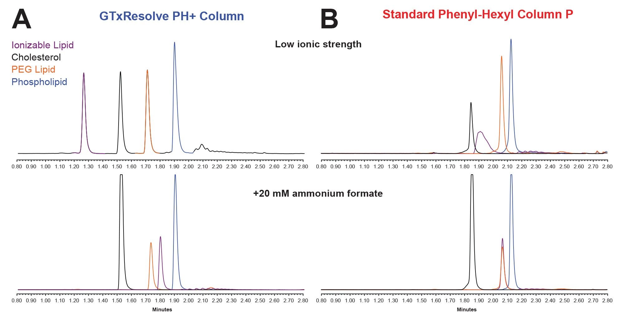
The reversed-phase chromatographic separation of lipids, which are inherently highly hydrophobic compounds, can be effectively realized on stationary phases featuring reduced hydrophobic retention. Previous column screenings identified the ACQUITY Premier CSH™ Phenyl-Hexyl Column as a suitable candidate with such characteristics.8 While the trifunctionally bonded C6-phenyl terminated ligand itself provides lower retention (roughly equivalent to C8 alkyl ligand), it is the charged surface that enables improved peak sharpness and symmetry for cationic analytes.9 GTxResolve RP 230 Å PH+ Columns were designed to build on this foundation and further refine lipid separations through: (i) increased surface charge density (enhancing performance for ionizable lipids), (ii) implementation of superficially porous particle architecture (improving mass transfer), and (iii) increased pore size (230 Å) to accommodate larger or partially associated lipid species. The activation of positive charge on the surface of GTxResolve PH+ Column occurs under acidic conditions (pH <5–6), which is best achieved using MS friendly volatile acids such as formic acid, even at concentrations as low as 0.1%.
The benefits of the charged surface are illustrated in Figure 1, which compares the elution characteristics of four representative lipids from commonly used classes (ionizable lipid: ALC-0315, cholesterol, PEG lipid: DMG-PEG 2000 and phospholipid: DSPC) under: a) low ionic strength conditions (0.1% formic acid) and b) in the presence of a salt additive (0.1% formic acid with 20 mM ammonium formate). For the GTxResolve Column, these conditions are sufficient to separate all analytes with symmetrical peak shapes (upper left). Conversely, such a low ionic strength mobile phase is inadequate for a standard phenyl-hexyl column (upper right), particularly for the ionizable lipid (purple trace), which exhibits poor peak shape consistent with insufficient surface charge. As this column was not specifically designed for lipid analytes, it displays low peak capacity (Pc = 8 vs 17 for GTxResolve Column at tg = 2 minutes), which does not improve with increasing ionic strength. Although addition of salt (lower chromatograms) corrects the peak shape of ALC-0315, the inherently limited selectivity prevents differentiation from the PEG lipid (right, orange vs purple), in contrast to the charged surface column. For the GTxResolve Column, increasing ionic strength primarily reduces repulsive electrostatic interactions, resulting in a corresponding increase in retention of the ionizable lipid (lower left). Notably, neutral analytes show minimal retention time shifts with changing ionic strength for both columns (cholesterol, PEG lipid, DSPC; black, orange, blue) highlighting a selective and tunable interaction mechanism specific to ionizable lipids. Such behavior can be conveniently exploited as a selectivity modulation parameter in method development.
Ionic Strength as a Selectivity Switch for Cationic Analytes
Since the retention of ionizable lipids can be adjusted independently of non-cationic components, the dependence of this effect as a function of ionic strength is further studied. Ammonium formate was used as a salt additive, and its concentration was varied between 0 mM and 20 mM. The retention of a permanently ionized lipid, DOTAP, as well as the components of the LNP2 formulation containing a proprietary hydrophobic lipid was mapped, and the results are shown in Figure 2A and 2B. The data clearly illustrates a considerable increase of retention times for both ionizable lipids, with the effect plateauing at concentrations >10 mM. The elution time of other components remains unchanged, and changes in retention times for both ionizable lipids can be described by a simple logarithmic function in the range of 0–10 mM. Both compounds are similarly affected by the increase in ionic strength, allowing for straightforward extrapolation of retention times from measurements acquired without salt additive.
Therefore, by measuring the retention time of an ionizable lipid in the absence of salt (RT0), the following empirical relationship can be applied:

This approach enables accurate prediction of retention times for other analytes, such as ALC-0315 (Figure 2C, yellow bars).
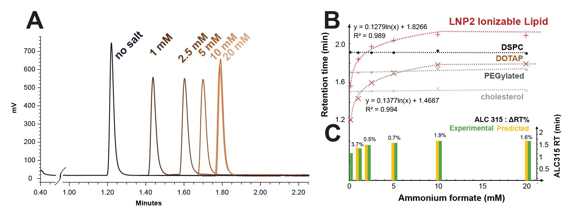
Experimental verification (green bars) revealed good agreement, with an average ΔRT = 0.017 minute (maximum ΔRT = 0.037 minute, corresponding to 3.7% for 1 mM).
Adjustment of ionic strength provides a simple and effective strategy to resolve co-elution between ionizable lipids and neutral components. In cases where lipids from different classes co-elute, alternative strategies, such as the use of organic mobile phase modifiers, may provide additional selectivity benefits.⁶
Eluent Additives for Peak Shape and Selectivity Improvement
PEGylated lipids often exhibit broader elution profiles compared to other lipid components, likely due to their inherent heterogeneity, which can negatively impact the overall analytical sensitivity. While steeper gradients lead to more pronounced peak sharpening, they generally will reduce the resolution between the PEG lipid and the closest eluting cholesterol peak. Isopropanol has previously been identified as an eluent additive that can improve the separation.8 While the GTxResolve PH+ Column delivers adequate resolution and sample throughput for most typical LNP formulations under generic conditions; the effect of a range of co-solvents is evaluated to determine if further improvements could be made on the PEG lipid peak shape. Volatile eluents, specifically alcohols, were the focus, by adding them to both mobile phases A and B at 10% (v/v); results are shown in Figure 3.
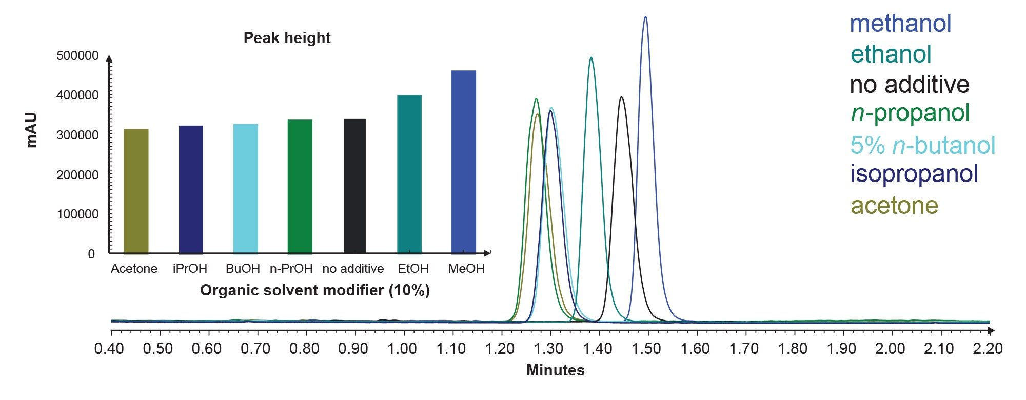
The screening results revealed superior performance for MeOH and EtOH containing mobile phases. Despite the slightly lower elution strength of the MeOH containing mobile phase, it yielded the sharpest peak (w50% = 0.037 minute vs 0.046 minute without MeOH, 25% improvement) and was therefore selected for further optimization. The result was verified by UV detection to exclude any potential ELSD-related matrix effects. The screening also demonstrated similar improvement for DSPC (w50% = 0.036 minute vs 0.045 minute without MeOH, 25% improvement). These findings suggest that the protic character of MeOH is important for improving peak shape of late eluting lipid components, likely by disrupting residual polar interactions with the stationary phase. This is further supported by the observation that more hydrophobic alcohols (propanols, butanol) as well as an aprotic solvent (acetone), did not provide comparable improvement.
To maximize the MeOH effect, further optimization was performed, establishing that 30% MeOH addition to mobile phase B only provided optimal performance. This condition delivered significant sharpening of the PEG lipid and DSPC peaks without compromising resolution between ionizable lipids (as observed when MeOH was added also to mobile phase A) or overall elution strength (as observed at MeOH concentrations >30%). Subsequently, the effect of different organic modifiers at a 30/70 ratio with MeCN (MeOH, EtOH, and IPA) was evaluated. The resulting chromatograms, illustrating differences in selectivity and retention, are shown in Figure 4.
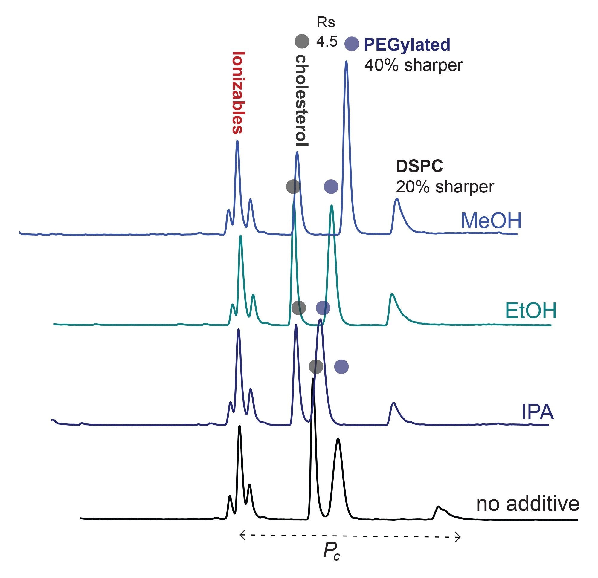
Significant peak sharpening of PEG lipid and DSPC was confirmed using MeOH (~40% and ~20% improvement, respectively). Interestingly, all tested lipids were resolved under high-throughput conditions; however, overall peak capacity decreased in the presence of organic modifiers (by approximately 20–30%). A pronounced effect on selectivity between cholesterol and PEG lipid was observed (RsMeOH = 4.5, RsEtOH = 3.2, RsIPA = 1.7, Rsno additive = 1.8), revealing a clear trend in which increasing hydrophobicity of the alcohol correlates with reduced selectivity between those species. These results establish a practical method development guideline favoring the use of MeOH as a mobile phase modifier, except in cases where maximizing peak capacity (e.g. resolving multiple ionizable lipids and their related impurities) is prioritized.
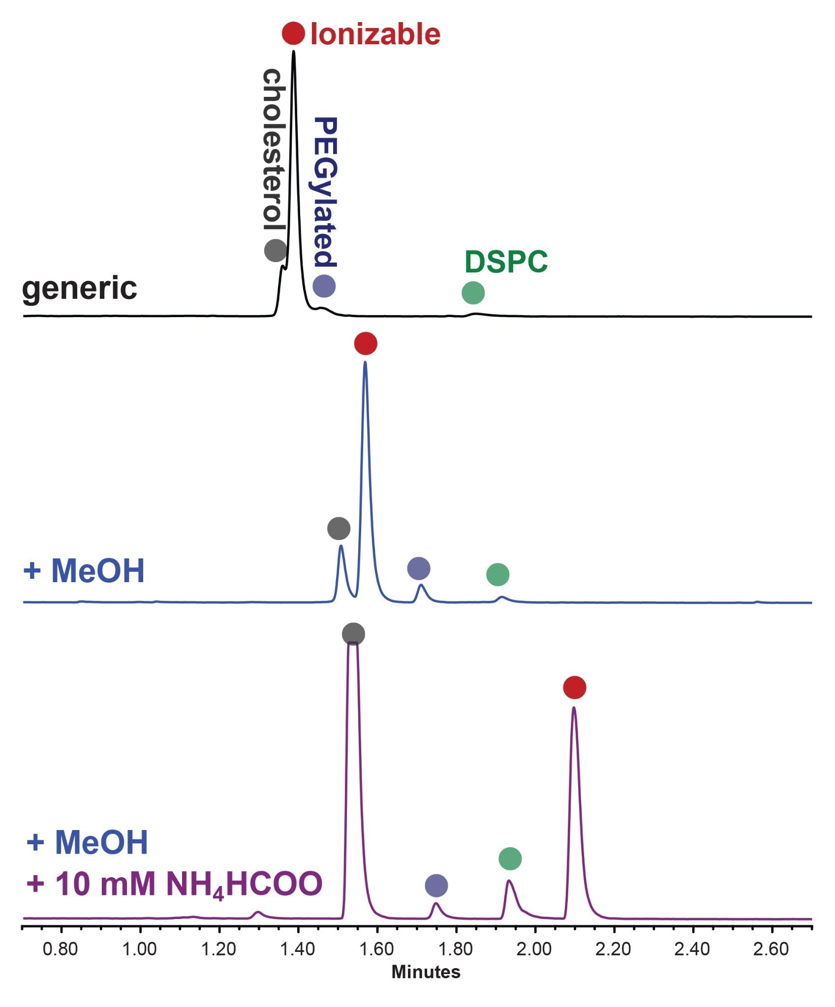
Separation of a Challenging LNP Formulation Following Established Guidelines
Finally, the separation of LNP2 was evaluated, containing a proprietary ionizable lipid with high hydrophobicity. When using generic high-throughput conditions with no additives, the separation was unsatisfactory, with partial co-elution of the ionizable lipid with both the PEG lipid and cholesterol (Figure 5). Applying MeOH as an MPB additive (blue trace) yielded the expected peak shape improvements and selectivity shifts, enabling the improved separation of the ionizable lipid. To further resolve the remaining partial co-elution with cholesterol (Rs = 1.8), 10 mM ammonium formate was introduced, resulting in full resolution of all four components (all Rs >3.3).
Notably, changes to mobile phase composition also impacted ELSD response, resulting in a significant increase in signal intensity, particularly for cholesterol and DSPC, providing additional sensitivity benefit. A detailed discussion of matrix effects and detection parameters influencing sensitivity is provided for ELSD10 and CAD11 detection. The approaches presented here provide practical, though non-exhaustive, guidance for LNP separation development. However, additional optimization opportunities remain. For instance, increasing the column temperature (e.g. from 40 °C to 50 °C) could globally reduce retention, and further refinement of the gradient profile may enable even shorter analysis times than the 4-minute method presented. On the other hand, analysis of negatively charged compounds (e.g. phospholipids) under conditions with activated surface likely requires increasing of the mobile phase ionic strength and column temperature to allow for efficient elution of those highly retained species.
Conclusion
This application note describes method development considerations for the compositional analysis of LNPs using GTxResolve RP 230 Å PH+ Columns. General guidance is provided for improving lipid separations by leveraging key column design features, including an acid-activated positively charged surface. This design enables straightforward interpretation of key method parameters, such as ionic strength, and facilitates rationalization of impact of mobile phase additives. The guidelines presented here are relevant for the described high-throughput conditions using short columns and are expected to be broadly applicable to lipid classes studied. In this work, high flow rates (1 mL/min) and short gradients (2 minutes) were employed to achieve rapid analyses (4 minutes total runtime), while still providing for effective selectivity optimization through the deliberate use of ionic strength and organic modifiers. Selectivity behavior may differ when using longer columns or alternative gradient profiles. For example, in lipidomics applications, longer gradients may be beneficial to increase peak capacity. Accordingly, flow rate, gradient time, and column length should be optimized collectively due to their interdependence. As with other reversed-phase separations, model-based optimization approaches using chromatographic software may further enhance method development efficiency. Ongoing work is focused on improving separations of structurally similar lipid impurities.
References
- L. Antonara; et al. A review, lipid-based drug delivery systems: concepts and recent advances in transdermal applications, Nanomaterials 15 (2025) 1326. https://doi.org/10.3390/nano15171326
- K. Sandra; A. dos Santos Pereira; G. Vanhoenacker; F. David; P. Sandra. Comprehensive blood plasma lipidomics by liquid chromatography/quadrupole time‑of‑flight mass spectrometry, J. Chromatogr. A 1217 (2010) 4087–4099. https://doi.org/10.1016/j.chroma.2010.02.039
- U.S. Food and Drug Administration, Guidance for Industry, Liposome Drug Products: Chemistry, Manufacturing, and Controls; Human Pharmacokinetics and Bioavailability; and Labeling Documentation, FDA, Silver Spring, MD, 2018. https://www.fda.gov/media/70837/download
- M. Ovčačíková; M. Lísa; E. Cífková; M. Holčapek. Retention behavior of lipids in reversed‑phase ultrahigh‑performance liquid chromatography–electrospray ionization mass spectrometry, J. Chromatogr. A 1450 (2016) 76–85. https://doi.org/10.1016/j.chroma.2016.04.082
- Y. Fan; M. Marioli; K. Zhang. A review, analytical characterization of liposomes and other lipid nanoparticles for drug delivery, J. Pharm. Biomed. Anal. 192 (2021) 113642. https://doi.org/10.1016/j.jpba.2020.113642
- G.J. Schad; S. Fujisaki. Streamlined method development for efficient and reliable lipid nanoparticle analysis, LC GC Eur. 21 (2025) 19–23, https://www.chromatographyonline.com/view/streamlined-method-development-for-efficient-and-reliable-lipid-nanoparticle-analysis
- M. Imiolek; S. M. Koza. High throughput LNP Compositional Analysis using GTxResolve™ RP 230 Å PH+ Columns: Method Development Considerations, Waters Application Note (2026). 720009424.
- B.A. Alden; G. Isaac; W. Chen; M.A. Lauber. Lipid nanoparticle compositional analysis using charged surface hybrid phenyl‑hexyl separation with evaporative light scattering detection, Waters Application Note (2021) 720007331.
- P.C. Iraneta; K.D. Wyndham; D.R. McCabe; T.H. Walter. A review of Waters hybrid particle technology. Part 3: Charged surface hybrid (CSH) technology and its use in liquid chromatography, Waters White Paper (2011) 720003929.
- K. DeLaney; D. Han; R.E. Birdsall; Y.Q. Yu. Optimized ELSD workflow for improved detection of lipid nanoparticle components, Waters Application Note (2022) 720007740.
- R. Birdsall; X. Du; P. Bigos; D. Han; N. Bhiwankar. Automating charged aerosol detection (CAD) analysis with Empower™ CDS software using a single‑vendor integrated LC platform, Waters Application Note (2026) 720009297.
Disclaimer
Waters, GTxResolve, ACQUITY UPLC, QuanRecovery, MaxPeak and CSH are trademarks of Waters Corporation or its affiliates. Spikevax is a trademark of ModernaTx, Inc.
Featured Products
720009424, June 2026