Deformulating Size Exclusion Chromatography for LNP Payload Quantitation
Abstract
This application note discusses the use of a GTxResolve™ Premier BEH™ SEC 450 Å Column for the online deformulation of LNPs and size exclusion separation of the mRNA payload. Common detergent disruption protocols have been tested and found to lead to incomplete deformulation of the sample, as both intact LNPs and freed nucleic acids can be still observed in the SEC chromatograms. While pursuing alternative options, we discovered that a detergent and organic solvent could be applied as mobile phase additives to achieve complete disruption of intact LNP samples upon their injection into the LC system, thus removing the need for a sample preparation step. With the developed method, nucleic acid payloads can be effectively liberated from the lipid vehicle, size separated and quantified in a rapid and robust assay requiring only a minimal amount of sample. Such an assay provides instant insights into an otherwise challenging characterization of LNP payloads and is especially suitable to samples with multiple payloads important for emerging gene editing therapies.
Benefits
- Fast (<10 minutes) and efficient deformulating SEC-UV method for an integrity check and quantitation of a nucleic acid payload released from its LNP carrier
- Facile assay (no sample preparation) that takes advantage of the denaturing power of an amphiphilic detergent and an organic solvent mobile phase additive
- Readily deployable (no optimization required) SEC-UV separation to provide a cost and time-effective means to comprehensively quantify and characterize multiple payloads in different LNPs
Introduction
Cell and gene therapy drugs are providing new chances to treat previously intractable diseases. For drug developers, the promise of their effectiveness comes with a formidable challenge of ensuring the safety and efficacy of increasingly complex therapeutics. These drugs are multicomponent ensembles (carrier + payloads), which means there is an array of critical quality attributes (CQAs) that must be monitored at each stage of their development – creating bottlenecks in the process of bringing a new drug product to market.1 High-throughput analytical methods based on chromatographic separations can help facilitate their development and potentially even reduce costs.
Quantification of nucleic acid content within a lipid nanoparticle (LNP) is one example of an assay that deserves more attention. While measuring the content at the drug substance level is relatively straightforward (UV absorption) the same measurement cannot be as simply performed on the drug product. LNPs, despite having no UV chromophores, produce a significant amount of UV spectrophotometer signal at a 260 nm wavelength, due to the scattering of incident light.2 As the phenomenon is affected by sample properties, it can be difficult to correct a UV absorbance for this light scattering effect.3 Other techniques can be applied but they might involve laborious sample preparation as is the case with capillary electrophoresis (CE), or they might not yet be supported by peer reviewed publications.4
Alternative measurements with advanced separation and detection methods can be applied but they require specific reference materials (e.g. empty LNPs for MALS) or they require the use of certain modes of denaturing chromatography.2 Denaturing RPLC has been demonstrated to quantify the total RNA content of LNPs samples. In a recent piece of work, a low retentivity method was applied and different payloads were quantified together in a co-eluting peak.2 Application of retention inducing ion pairing reagents in the mobile phase offers the possibility to distinguish between different nucleic acids. However, use of amphiphilic detergents needed for complete disruption of the LNPs is not compatible with ion pairing RPLC stationary phases and methods. If loaded onto a column, they can cause poor reproducibility of the method, interference peaks and short lifetime of used columns.
Size exclusion chromatography has become a favored approach for the characterization of several emerging new modalities (AAVs, nucleic acids, mRNA/LNPs etc.).5–6 Significant attention is being placed on creating fit-for-purpose SEC methods for lipid nanoparticles (LNPs) and their payload drug substances. Optimized widepore, low adsorption SEC columns (>450 Å) are being developed such that new techniques can be envisaged. Past investigators have applied a positive surface potential gel permeation resin to perform LNP size exclusion chromatography.7 There is risk to this approach due to the potential adsorption and incomplete elution of free nucleic acid impurities. As such, we have alternatively investigated non-ionic, hydrophilic packing materials for SEC of LNPs and nucleic acids.
We hypothesized that quantification of LNP payload can be performed with a purpose-built SEC column if suitable disruptive sample preparation and chromatography conditions could be devised. As such, we have herein developed a method of online deformulation of LNPs using a GTxResolve Premier BEH SEC 450 Å SEC Column along with an optimized denaturing mobile phase. In this application note we optimized a deformulating mobile phase with an amphiphilic detergent (sodium dodecyl sulfate) together with an organic solvent (isopropanol) to achieve complete disruption of the LNPs along with an immediate SEC separation of the liberated nucleic acid payloads. The conditions of the SEC mobile phase quantitatively produce nucleic acid analytes that are free of any appreciable quantities of ion pairing, ionizable lipid or residual nanoparticle structures.
Experimental
The chemicals and solvents used in the study were used as received (P3813, phosphate buffered saline pouches, Sigma-Aldrich, Water Fisher Chemical, isopropanol Optima LC-MS - IPA (10684355, Fisher Chemical), water UHPLC gradient grade, 11357090, Fisher Chemical, Sodium dodecyl sulfate - SDS, L3771, Sigma-Aldrich, Triton™ X-100 Detergent, X100, Sigma-Aldrich). The LNP samples were transferred to QuanRecovery™ Vials low adsorption vials (Waters™ p/n: 186009186, Waters Corporation, Milford, MA) and used as received. Moderna COVID-19 vaccine (NDC 80777-279-99, Spikevax™ (COVID-19 Vaccine, mRNA), nominal mRNA concentration 0.1 mg/mL), Pfizer COVID-19 vaccine (NDC 59267-1025-4, Comirnaty™ (COVID-19 Vaccine, mRNA), nominal mRNA concentration 0.1 mg/mL), and PackGene™ Firefly Luciferase-mRNA (Cap 1+N1meψUTP, SM-102, nominal mRNA concentration 0.2 mg/mL) LNPs were used. As a reference material eSpCas9(N1) mRNA (230807A, GenScript) was used. PBS mobile phase was filtered before use (0.2 µm membrane, PES, 5660020, Nalgene). No sinkers were used at the end of lines and the instrument was periodically flushed (weekly or more frequently) with 70% isopropanol solution to prevent microbial contamination.
Alcohol mediated precipitation was performed to obtain reference mRNA material. Briefly, 50 µL of the LNP sample was diluted with 1 mL of 60 mM NH4OAc in 100% IPA, which causes the encapsulated mRNA to precipitate. The samples were spinned down (14,000 xg at 4 °C, 15 minutes), supernatant was removed and the pellet was washed with additional 1 mL of IPA. The samples were spinned down again, supernatant removed and the remaining pellet was dried in a N2 stream (5 minutes) and dissolved in 50 µL of water. This protocol was adapted from literature.8
LC Conditions
|
LC system: |
ACQUITY™ UPLC™ H-Class PLUS Bio System (quaternary) |
|
Detection: |
ACQUITY UPLC TUV Detector with 5 mm titanium flow cell, 260 nm and 230 nm; (2 points/s) |
|
Vials: |
QuanRecovery with MaxPeak™ HPS 12 x 32 mm Screw Neck Vial, 300 µL, 100/pk, (p/n: 186009186) |
|
Column: |
GTxResolve Premier BEH 450 Å SEC, 2.5 µm, 4.6 x 150 mm Column (p/n:186010584) |
|
Column temperature: |
40 °C |
|
Sample temperature: |
6 °C |
|
Injection volume: |
0.1–1.0 µL |
|
Flow rate: |
0.25–0.5 mL/min |
|
Mobile phase: |
1X PBS ie. (137 mM NaCl, 2.7 mM KCl, 10 mM Na2HPO4, 1.8 mM KH2PO4) pH 7.4, 20% isopropanol, 0.2% sodium dodecyl sulfate, 0.2 µm sterile filtered |
Results and Discussion
Native SEC Allows Fractionation of Intact LNPs
SEC separations are usually run under native conditions using aqueous buffers closely resembling physiological conditions. When applying such a condition to LNPs, there is a chance for analysts to observe some intact sample components, even if they are predominantly excluded from the column pores (Figure 1A). Developing a fit for purpose SEC-UV method for intact LNPs takes many considerations and careful optimization of mobile phases and packing materials, potentially having an average pore diameter as large 2000 Å. With the result obtained on a GTxResolve Premier BEH SEC 450 Å Column with 1X PBS mobile phase, it cannot be assured that the recovery of intact LNP is unbiased, reproducible, or quantitative. The goal of this work is to achieve a deformulating analysis, but the method development considerations start with an assessment of intact LNPs and detecting remnants of their intactness.
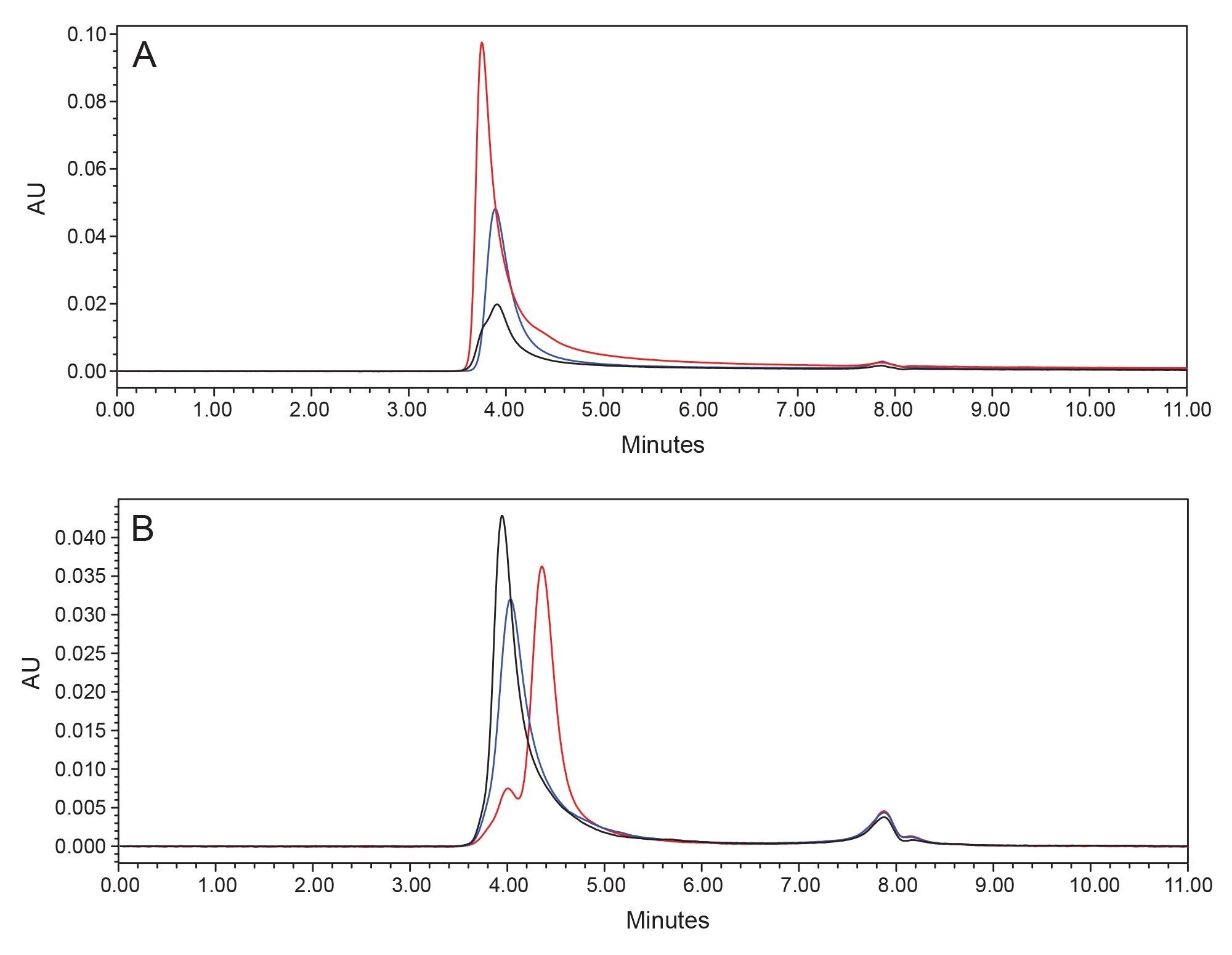 Figure 1. Overlay of non-denaturing SEC-UV (1X PBS, 25 °C) chromatograms of A) tested LNP samples B) their extracted mRNA payloads (spike protein mRNA: black Spikevax (COVID-19 Vaccine, mRNA), blue Comirnaty (COVID-19 Vaccine, mRNA), FLuc mRNA red PackGene LNPs).
The similarity of size of tested LNPs (SpikeVax (COVID-19 Vaccine, mRNA), Comirnaty (COVID-19 Vaccine, mRNA) around 80 nm and PackGene LNPs around 100 nm) is reflected in their close elution times, which were observed to range from 3.75 minutes to 3.91 minutes. This is despite the fact the cargo mRNA differs fairly significantly between each sample.
Both Comirnaty (COVID-19 Vaccine, mRNA) and SpikeVax (COVID-19 Vaccine, mRNA) COVID vaccines contain a construct of SARS-CoV-2 spike protein mRNA (around 5000 nt) while the PackGene LNPs were formulated with a smaller firefly luciferase (Fluc) mRNA (around 2000 nt). For a comparative analysis, the encapsulated mRNA from these LNP samples were extracted via isopropanol precipitation then analyzed with native SEC. It was found that the spike protein and firefly luciferase mRNAs could be separated from each other (4 minutes versuss 4.3 minutes, Figure 1B). Additionally, analysis of absorption at different wavelengths could be performed to further characterize the eluting species. It is well established that pure RNA has a characteristic ratio of absorbance at 260 versus 230 nm of 1.8–2.2.9 Indeed, when calculated for the reference material, the ratio was close to two. For the intact LNPs, the dominant signal was detected at 230 nm (since scattering effects are stronger at shorter wavelengths).10 A comparison of different parameters is provided in Table 1. Altogether, these data show that a standard SEC analysis performed under native conditions does not lead to the disruption of the LNPs. If a deformulation analysis is to be performed, additional considerations must be made.
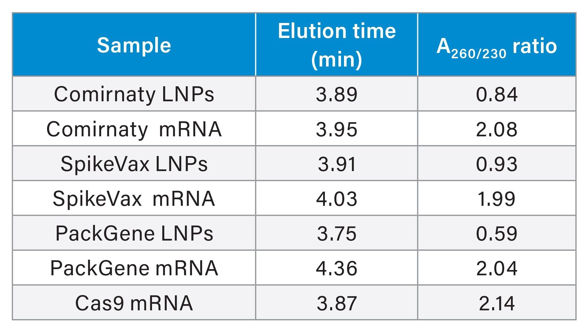 Table 1. Elution time and absorbance ratio parameters for different species obtained during native SEC analysis.
Standard Disruption Protocols Lead to Incomplete Deformulation of LNPs
mRNA content of the LNPs can be released by dilution with various denaturing agents, the most common being the non-ionic surfactant Triton X-100 Detergent.11 We evaluated such conditions by first diluting samples to 0.1% final detergent concentration for the disruption of the LNPs. It was found that such a treatment and subsequent native SEC separation was not sufficient to achieve complete disruption as evidenced by inconsistent A260/230 ratios and a residual LNP peak, which was most easily observed for PackGene LNPs (Figure 2). Due to the overlapping elution times of COVID mRNAs and intact LNPs, we found A260/230 ratios to be the best parameter to monitor completeness of denaturation/deformulation.
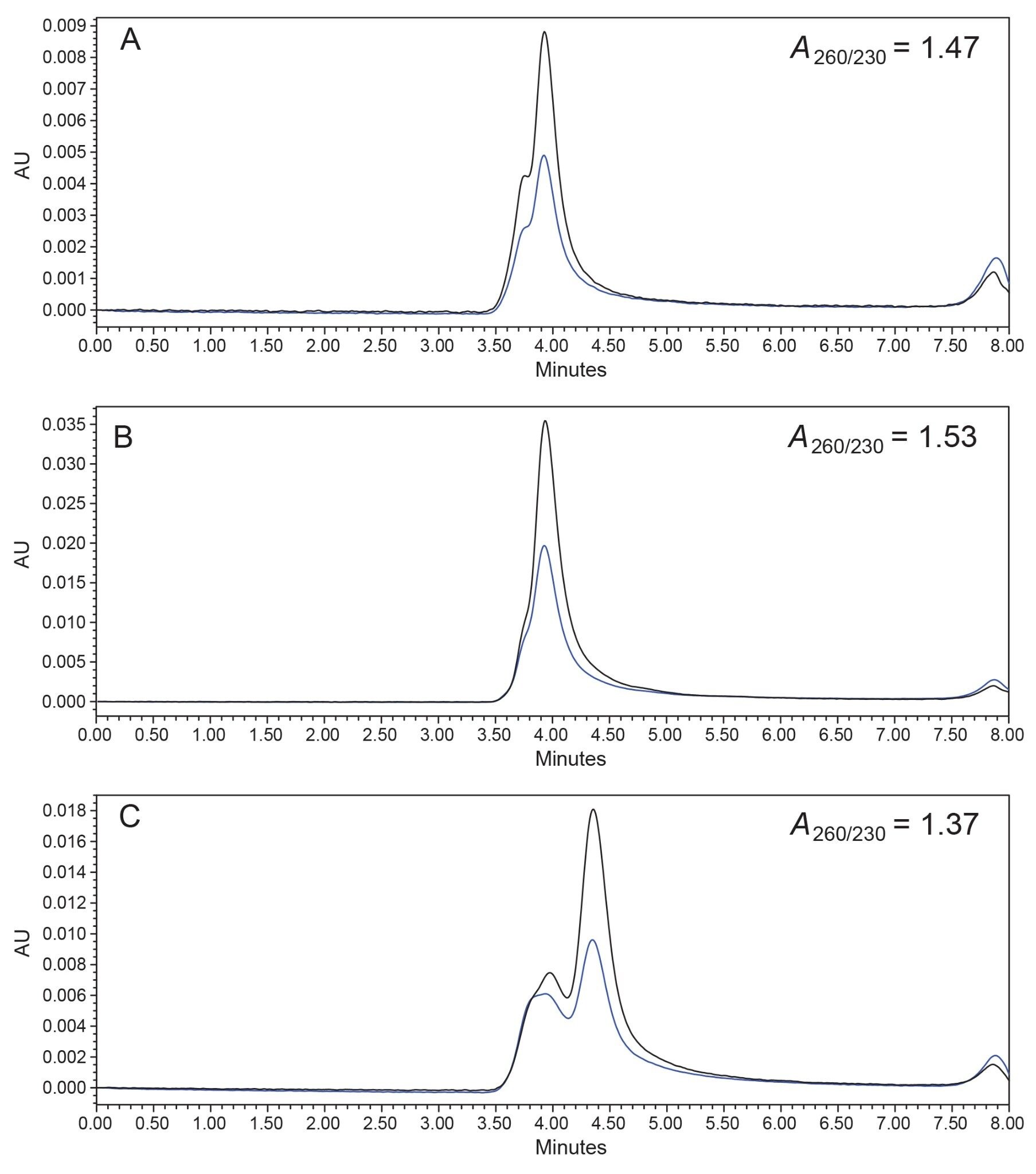 Figure 2. Overlay of native SEC-UV (1X PBS, 25 °C) chromatograms (black 260 nm, blue 230 nm) of Triton X Detergent disrupted LNP samples A) SpikeVax (COVID-19 Vaccine, mRNA), B) Comirnaty (COVID-19 Vaccine, mRNA), C) PackGene LNPs along with the calculated A260/230 ratios. All samples were diluted to 0.1% Triton X-100 Detergent concentration before injection.
Consequently, a 0.1% Triton X-100 Detergent offline deformulation was confirmed to be insufficient. Stronger concentrations could have been pursued but it was additionally found that residual Triton X-100 Surfactant (which absorbs at 260 nm) is retained past the void column volume and elutes in a tailing peak that might influence subsequent analysis. Further attempts to optimize conditions (temperature, surfactant content, etc.) were not fruitful. We were unable to find sample pre-treatment steps that could be combined with a non-denaturing SEC method to produce fully denatured LNP formulations.
Complete and Universal Deformulation With an SDS and IPA Containing SEC Mobile Phase
Exhaustive screening of conditions that enable denaturation of the sample (as judged by A260/230 ratios reaching value of 2) allowed us to identify the key role of mobile phase additives. When 0.2% SDS and 20% IPA were included in the mobile phase, it was found that LNPs could be universally deformulated without the need for additional sample manipulation (leading to expected A260/230 ratios). Raising the temperature of the column to 40 °C was found to be beneficial to improving the recovery of the payload analytes and reducing the pressure drop across the column.
We used SDS instead of Triton X-100 Detergent, as this anionic detergent is transparent at 260 nm wavelength of interest. IPA was essential to completing the deformulation and increasing the solubility of SDS. We verified the completeness of disruption by further raising the column temperature to 55 °C and the SDS concentration to 1.0%. Comparable results were obtained, suggesting that 40 °C and 0.2% are sufficiently high parameters. Increased amounts of detergent and higher column temperatures may lead to decreased column lifetime, so it is a balance of considerations that led us to finalize on 40 °C and 0.2% method conditions.
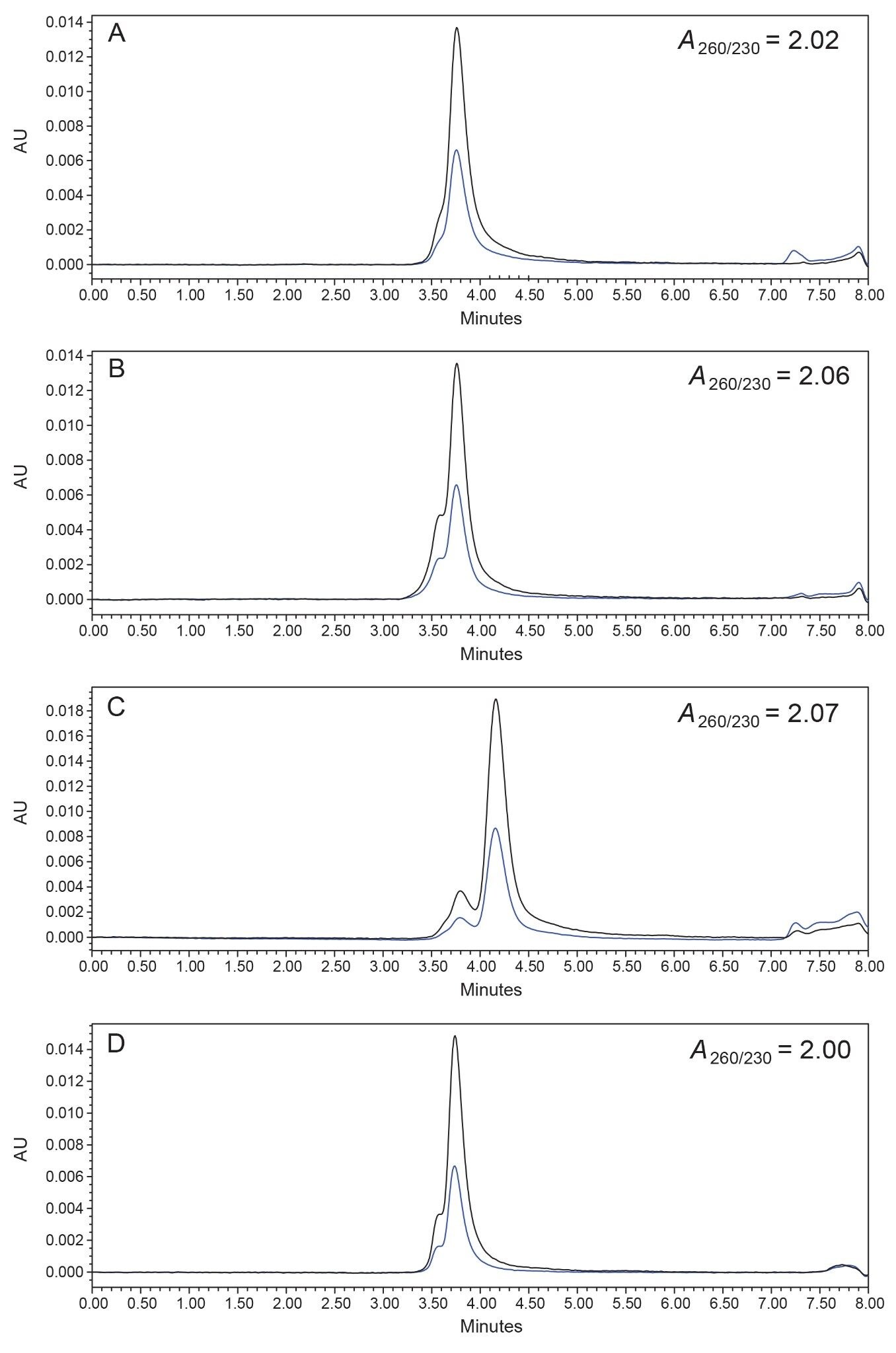 Figure 3. Overlay of deformulating SEC-UV (1X PBS 20% IPA, 0.2% SDS, 40 °C) chromatograms (black 260 nm, blue 230 nm) of intact LNP samples A) SpikeVax (COVID-19 Vaccine, mRNA), B) Comirnaty (COVID-19 Vaccine, mRNA), C) PackGene LNPs as well as D) Cas9 mRNA reference material along with their A260/230 ratios.
It is important to notice that SEC enables the separation of different size mRNA payloads as exemplified by the different elution times of FLuc mRNA and Spike protein mRNA (4.16 minutes versus 3.76 minutes, Figure 4) as well as analysis of aggregation profiles (pre-peaks). High resolution separation of such similarly sized payloads would require optimization of conditions or the use of a column packed with a material of a different pore size, such as the GTxResolve Premier SEC 1000 Å Column.12 However, the GTxResolve Premier BEH SEC 450 Å Column is purposely proposed here to facilitate fast abundance determinations of a Cas9 mRNA (5000 nt) and guide RNA (100 nt) – two example drug substances used in dual payload LNPs for gene editing.
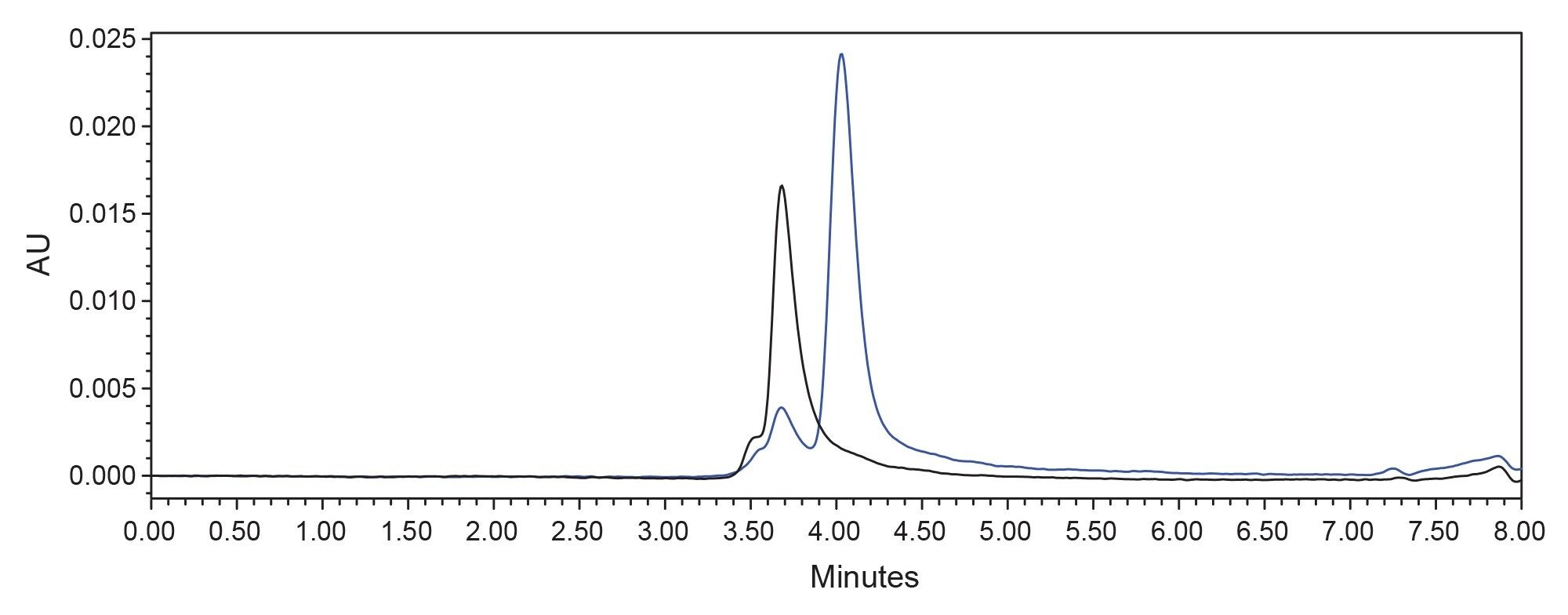 Figure 4. Overlay of deformulating SEC-UV (1X PBS 20% IPA, 0.2% SDS, 40 °C) 260 nm chromatograms for intact LNP samples containing mRNA of different size spike protein mRNA (Spikevax LNPs, black) or FLuc mRNA (PackGene LNPs, blue).
Comparison of the peak areas of different LNPs and a reference material, given their known concentrations, show similar response suggesting full recovery of the nucleic acid components (Figure 5). This is consistent with the fact that RNA of similar mass concentrations should yield similar signal (A260=1 at 40 µg/mL).10 Note that every mRNA molecule will exhibit a unique extinction coefficient as well as recovery under analysis conditions. An exact match reference material should thus be used to achieve accurate and precise quantification.
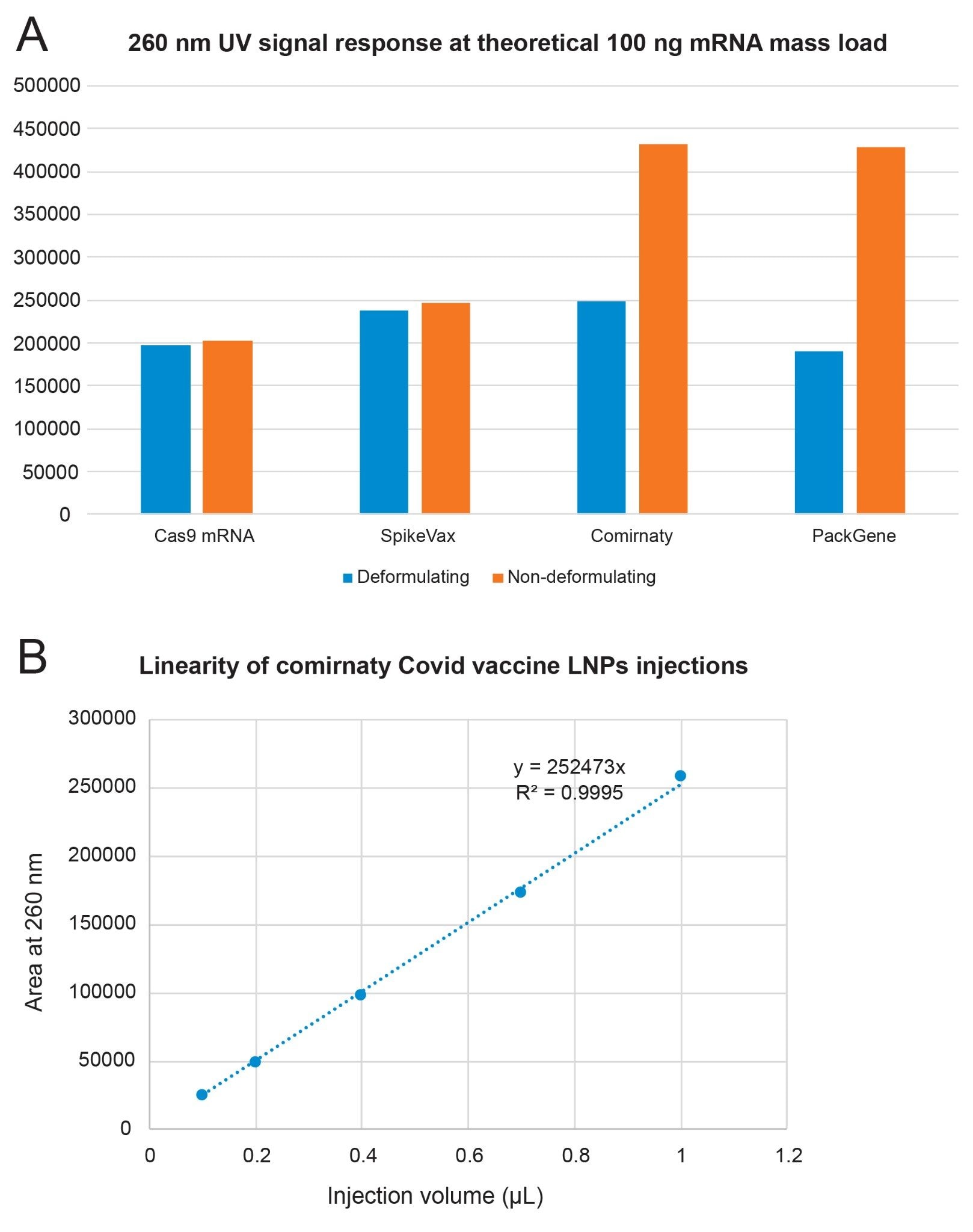 Figure 5. A) Analysis of absolute peak areas at 260 nm for different LNPs samples and reference mRNA material under deformulating SEC-UV (blue) and native SEC conditions (orange) B) Linearity of detector response when injecting different amounts of Comirnaty LNPs.
Conclusion
In this application note, we have shown that efficient size-based separation of a nucleic payload is possible. In addition to its utility for mRNA vaccines, the corresponding method will enable quantitation of gene editing LNPs containing both Cas-type mRNA and CRISPR gRNA. Various agents and LNP formulations were investigated to ensure universality of the disrupting conditions and the development of a platform method. We hypothesize that the presented conditions could be applicable to analyzing the payload of a protein nanoparticle, wherein a nucleic acid is encapsulated by a recombinantly expressed protein or an engineered virus.
In summary, we demonstrate the use of a new deformulating SEC-UV method to release mRNA content from an LNP to allow a quantitative analysis of the payload material. This method does not require sample preparation and it is based on the use of a deformulating mobile phase comprised of an amphiphilic detergent and an organic solvent. Importantly, SEC provides a means to separate different payloads, which is essential for emerging gene editing therapies which deliver both Cas mRNA and a guide RNA in the same drug product.
References
- Sparmann, A., & Vogel, J. EMBO J, 42(21), e114760. 2023.
- Jia, X., Liu, Y., Wagner, A. M., Chen, M., Zhao, Y., Smith, K. J., ... & Pennington, J. J Chrom B, 1186, 123015. 2021.
- Porterfield, J. Z., & Zlotnick, A. A. Virology, 407(2), 281–288. 2010.
- SCIEX Application Note https://sciex.com/tech-notes/biopharma/mrna-lnp-nucleic-acid-assessment-from-distinct-formulations-by-m.
- Fekete, S., Aebischer, M. K., Imiołek, M., Graf, T., Ruppert, R., Lauber, M., … & Guillarme, D. TrAC, Trends Anal. Chem., 117088. 2023.
- Fekete, S., Doneanu, C., Addepalli, B., Gaye, M., Nguyen, J., Alden, B., ... & Lauber, M. J Pharm Biomed Anal, 224, 115174. 2023.
- Zhang, J., Haas, R. M., & Leone, A. M. Anal chem, 84(14), 6088–6096. 2012.
- Packer, M., Gyawali, D., Yerabolu, R. et al. Nat Commun, 12, 6777. 2021.
- Koetsier, G. & Cantor, E., New England Biolabs Technical Note, (2019), 7/19 https://www.neb.com/en/-/media/nebus/files/application-notes/technote_mvs_analysis_of_nucleic_acid_concentration_and_purity.pdf?rev=c24cea043416420d84fb6bf7b554dbbb.
- Ramirez-Cuevas, (…), & Papakonstantinou, I. ACS photonics, 9(2), 672–681. 2022.
- Muramatsu, H., Lam, K., Bajusz, C., Laczkó, D., Karikó, K., Schreiner, P., ... & Pardi, N. Mol. Ther. 30(5), 1941–1951. 2022.
- D’Atri V. Lardeux H., Goyon A., Imiołek M., Fekete S., Lauber M., Zhang K., Guillarme D., Int. J. Mol. Sci. 25, 6254. 2024. https://doi.org/10.3390/ijms25116254.
720008549, October 2024