
DESI Mass Spectrometry Imaging of Soft Fruit
Wei Rao, Lisa Towers, Mark Towers, Joanne Ballantyne, Gemma Molyneux
Waters Corporation, United Kingdom
Published on September 04, 2026

這是一篇應用簡報,不含詳細的實驗內容章節。
Abstract
This application brief describes the use of desorption electrospray ionization (DESI) for the mass spectrometry imaging (MSI) of the high-water content fruits strawberries and raspberries. The use of three different direct transfer substrates to assess the compound transference and spatial changes are described when this method is utilized for soft fruit analysis.
Benefits
- Simple and quick to use
- Maintains mass spectrometer cleanliness
- Removes the requirement for challenging cryosection sample preparation
- Each transfer substrate has its benefits allowing application tailoring
- Identification of different substrates which preserve spatial localization and metabolite content during transfer
Introduction
Performing an MSI experiment on fruits or vegetables with a high water content can be difficult as the samples may be crushed or splintered during cryo-sectioning, leading to the loss of valuable localization information. Fresh samples may be cut with a knife and placed on the DESI surface as slices; however, this may lead to analytical challenges due to the softness of the tissue causing distortions during analysis, (which can be especially acute for sweet fruit samples) and potential for blockages in the inlet capillary. The mass spectrometer risks contamination from the high sucrose levels which can caramelize on the source surfaces and potentially block the DESI inlet tube. The intense sucrose signals can also lead to competitive ionization and detector saturation, reducing the detection of other analytes of interest.
To resolve these potential problems, it can be preferable to perform a direct transfer method, whereby the fruit or vegetable is cut using a knife and then imprinted onto a suitable substrate. This transfers the analytes from the sample onto the medium and the imprint is then imaged. The method has the benefit of not requiring the sample to be sectioned and also reduces the amount of analytes available for ionization, keeping the mass spectrometer and inlet cleaner.
The three transfer substrates being assessed for this application are glass microscope slides, commercially available nitrocellulose membranes, and Hamamatzu Poropare™ plates.
Results and Discussion
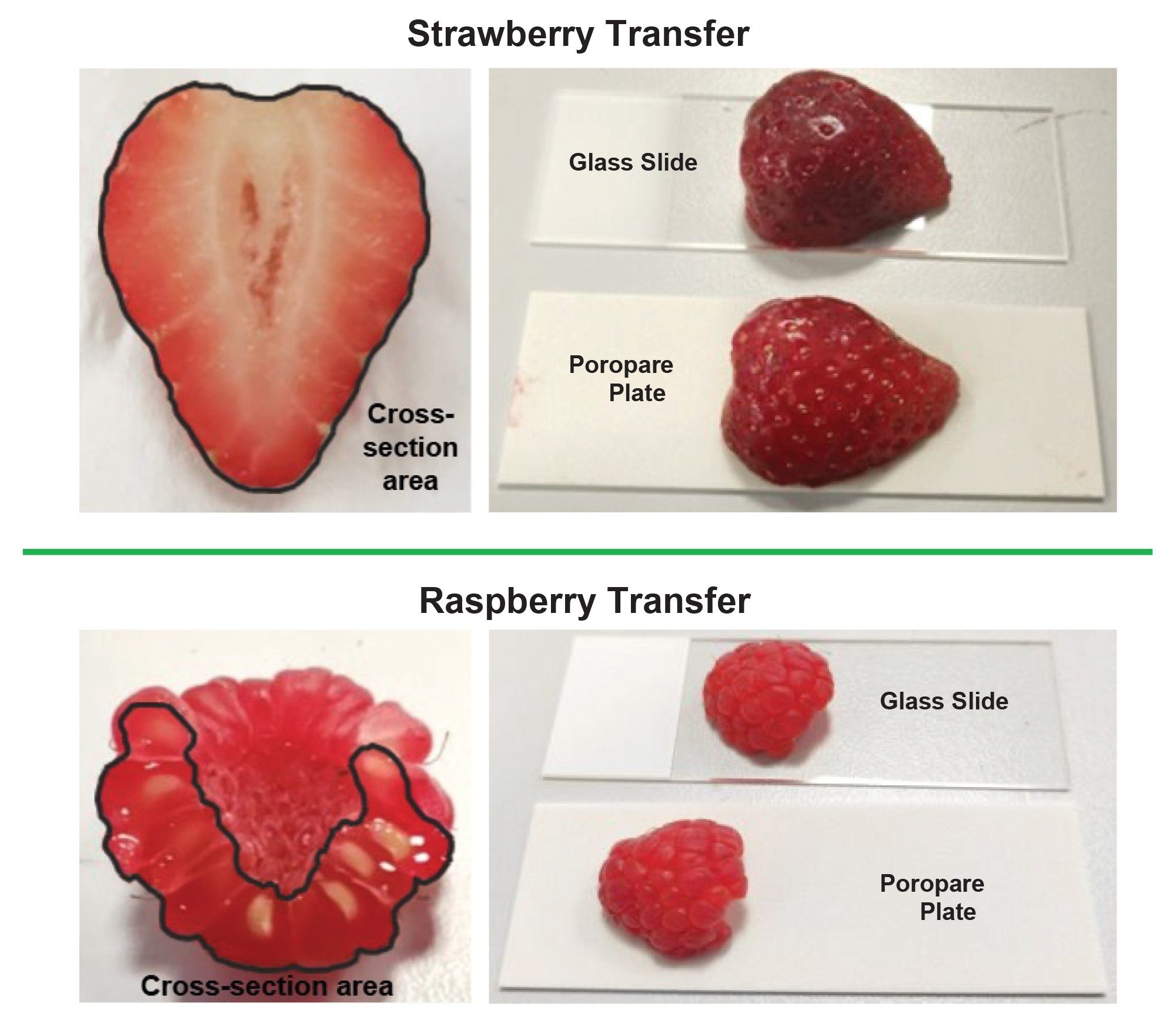
Fresh strawberries and raspberries were sourced from a local supermarket and cut with a knife directly through the center to expose a cross-section of the fruit. The cut surface of the fruit was then placed onto one of three transfer substrates and held in place for 10 seconds, allowing metabolites to transfer onto the medium. Figure 1 shows an example cross section and transfer of a strawberry and a raspberry onto a glass microscope slide and a Poropare plate.
Once transfer has been completed, the imprints of the fruits were allowed to air dry prior to analysis on a Xevo™ MRT P10 Mass Spectrometer coupled to a Waters DESI XS Source. MS imaging was performed in negative ionization mode, with a 50 µm pixel size and used a 95:5 MeOH:H2O spray solvent. The data was processed within Waters High Definition Imaging (HDI™) Software and lockmass correction was performed using the endogenous analyte sucrose at m/z 341.1089.
The nitrocellulose membrane proved fragile, possibly due to the high methanol content of the DESI solvent spray. The integrity of the membrane was compromised after just one experimental run, suggesting that this medium may only be suitable for a single DESI analysis per transferred section. In contrast, the glass microscope slide and Poropare plate both appeared to be robust when analyzed by DESI, potentially allowing for multiple passes should any material remain on the surface post primary analysis.
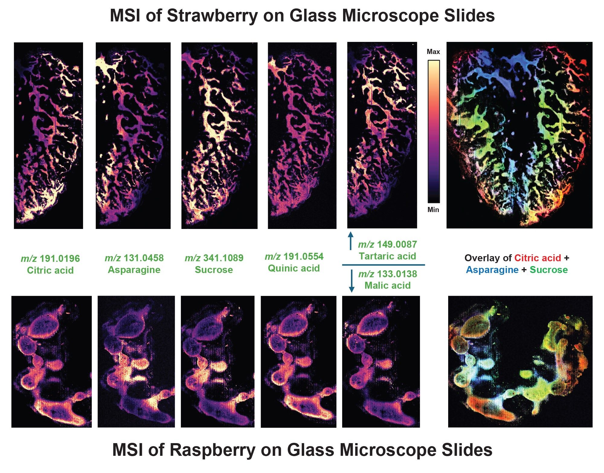
When the fruit slices were placed onto the glass microscope slides pooling of the transferred liquids from the samples were observed after the fruits were lifted off the slides. The MS images generated also indicated a high degree of delocalization of the detected metabolites (Figure 2). Analytes were generally detected with high signal intensities. Ion images were generated from analytes putatively assigned as: citric acid with a 0.13 mDa mass accuracy, asparagine with a 0.42 mDa mass accuracy and sucrose *endogenous lockmass, quinic acid with a 0.71 mDa mass accuracy, tartaric acid (strawberry) with a 0.46 mDa mass accuracy and malic acid (raspberry) with a 0.45 mDa mass accuracy. A red, green, blue (RGB) overlay was produced from three analytes (citric acid, asparagine, and sucrose), showing their relative differences in localization within the fruit impressions.
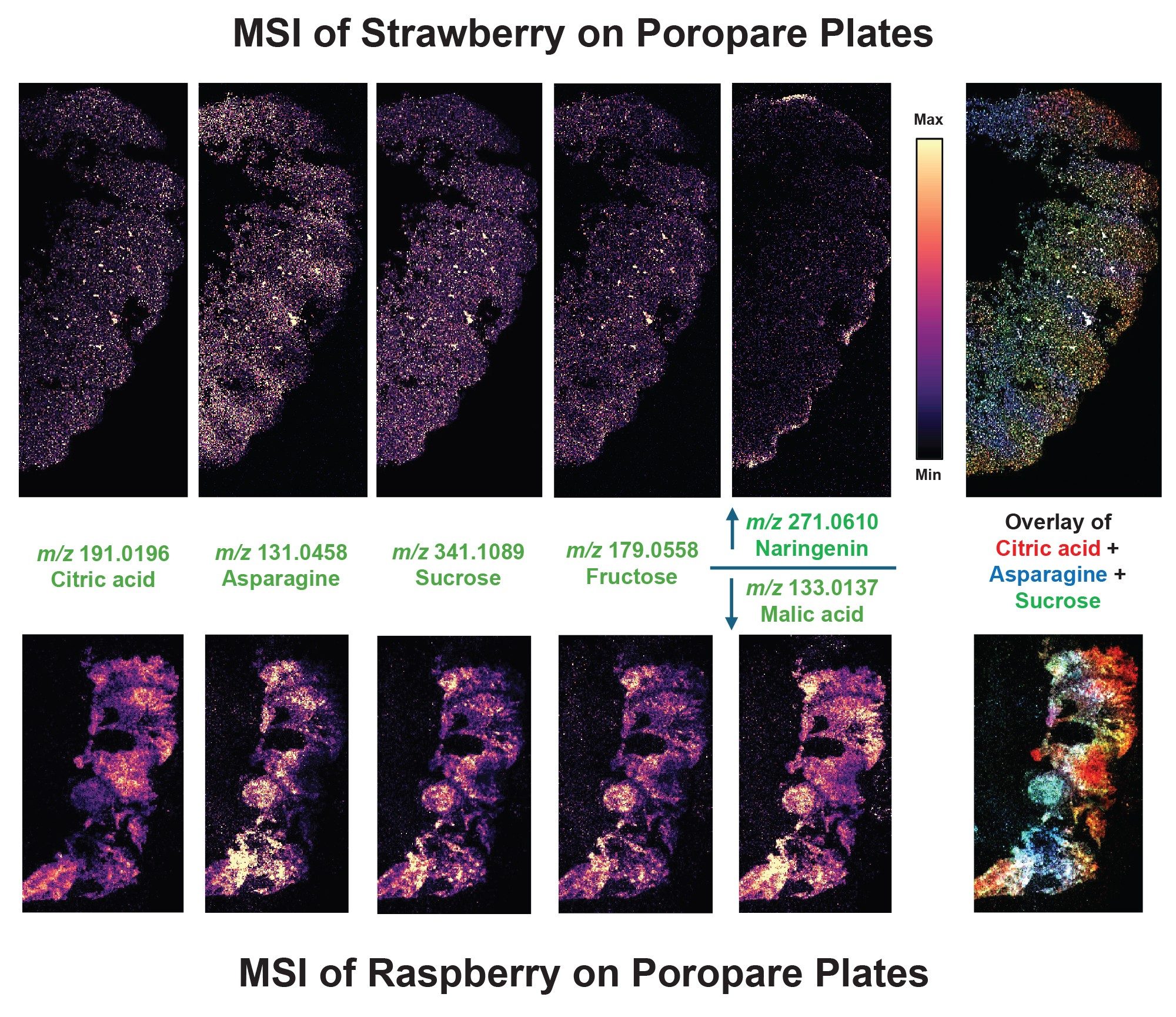
The metabolites from the fruit slices transferred the most effectively (for MSI) onto the Poropare plates. There was less delocalization observed compared to the other two substrates and features within both the strawberry and raspberry samples were resolved with good detail by the DESI MSI analysis. The segments within the strawberry and individual globules of the raspberry can be easily distinguished, particularly within the RGB overlay ion images (Figure 3). However, the analyte intensity was lower compared to the glass slides and the nitrocellulose membrane acquisitions. Ion images were generated from analytes putatively assigned as: citric acid with a 0.13 mDa mass accuracy, asparagine with a 0.52 mDa mass accuracy and sucrose *endogenous lockmass, fructose with a 0.31 mDa mass accuracy, naringenin (strawberry) with a 0.20 mDa mass accuracy and malic acid (raspberry) with a 0.45 mDa mass accuracy. An RGB overlay was produced from three analytes (citric acid, asparagine, and sucrose), showing their relative differences in localization within the fruit impressions.
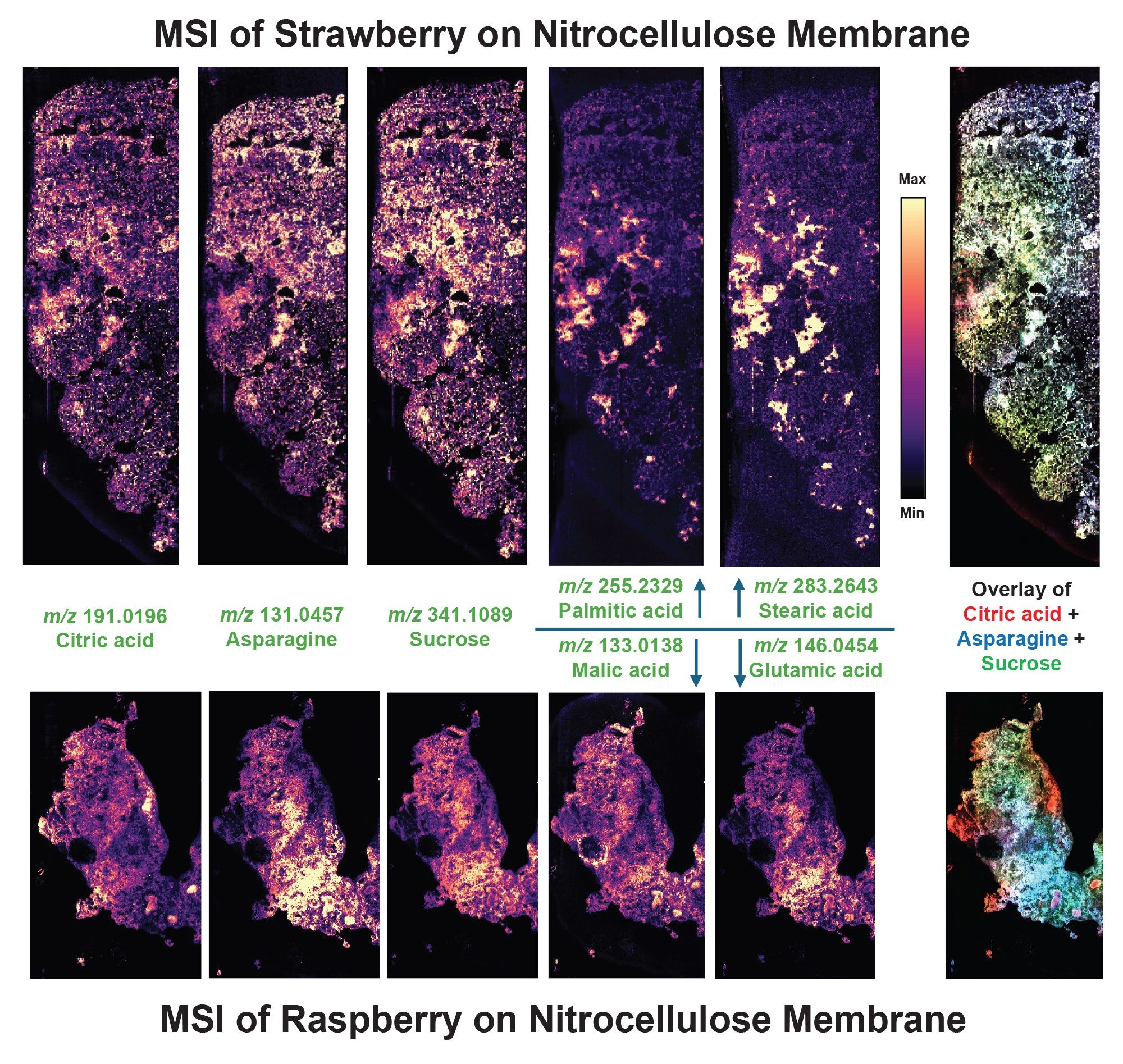
The fruit slice metabolites transferred well onto the nitrocellulose membrane with significantly less pooling and delocalization than can be seen with the glass microscope slides. A degree of delocalization can be observed compared to the transfer for the Poropare plates, as there were no globule sections observed for the raspberry sample and no distinct segments observed within the strawberry sample (Figure 4). Ion images were generated from analytes putatively assigned as: citric acid with a 0.13 mDa mass accuracy, asparagine with a 0.42 mDa mass accuracy and sucrose *endogenous lockmass, palmitic acid (strawberry) with a 0.05 mDa mass accuracy, stearic acid (strawberry) with a 0.05 mDa mass accuracy, malic acid (raspberry) with a 0.45 mDa mass accuracy and glutamic acid (raspberry) with a 0.48 mDa mass accuracy. An RGB overlay was produced from three analytes (citric acid, asparagine and sucrose), showing their relative differences in localization within the fruit impressions.
Conclusion
Utilizing a transfer substrate for metabolite transfer, DESI MS imaging of soft fruit samples was tested with three different substrates: glass microscope slides, nitrocellulose membranes, and Poropare plates from Hamamatzu. All three substrates took imprints of the metabolites from the soft fruit samples and produced ion images that were visualized within the HDI Software. The analyte signals were the strongest from the glass microscope slides; however, the highest level of metabolite delocalization was observed within this substrate. The Poropare plates demonstrated the most optimal preservation of metabolite localization out of all of the substrates compared; however, it also showed the lowest analyte intensity response amongst the substrates tested. The nitrocellulose membrane displayed a moderate loss of spatial localization during the transfer process; however, the substrate showed an improvement in analyte intensity compared to the Poropare membrane. Overall, the DESI XS platform was shown to be well suited for the MS imaging of soft fruit samples using a variety of different direct transfer substrates.
720009564, September 2026


