Aura+ System
Fast, decisive, low-volume particle characterization analysis for biotherapeutic workflows
When you need the flexibility to conduct particle characterization analyses on different types of biologics, including protein and antibody therapeutics, cell therapies, and gene therapies, the Aura+ System has you covered.
Fully configured to support applications across protein, antibody, cell, and gene therapy workflows, Aura+ brings particle characterization to earlier stages of therapeutic development, so you can make better decisions sooner.
Powered by the backgrounded membrane imaging (BMI) and fluorescence membrane microscopy (FMM) technology shared within the Aura family of instruments, Aura+ System enables applications from late discovery to development and manufacturing, enhancing decision-making and accelerating your success.
 Aura+ System
Specifications
|
Imaging area: |
24.6 mm2 (96-well); 158.9 mm2 (24-well) |
|
Optics: |
4x and 20x objectives |
|
Minimum volume: |
5 µL (assay dependent) |
|
Resolution: |
1.0 pixel/µm |
|
Detectable size range: |
From >1 µm (ECD) to <5 mm (ECD) on 96-well; <14 mm (ECD) on 24-well |
|
BMI read time: |
1 minute/sample |
|
FMM read time: |
30 seconds/sample |
|
Software: |
Particle VUETM 5.x all-in-one software suite for image and analysis (Security Pack available) |
Overview
- Save precious material with just 5 µL sample volume requirements
- Supports large sample volumes of 500 µL or more
- Get actionable results from a full 96 well plate of labeled samples to data in just 90 minutes
- Boost efficiency with 96-well and 24-well membrane plate high-throughput capability
- Gain deeper particle insights with advanced imaging
- Rely only on one platform for proteins, antibodies, and cell and gene therapies development
Recommended Use: For workflows requiring complete flexibility across a full spectrum of analytes, including biologics, viral vectors, cells, and other contaminants.
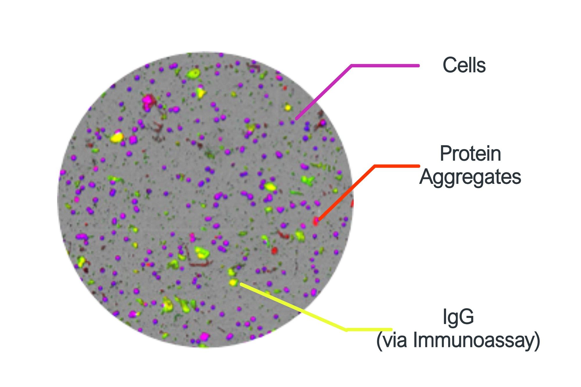
Take the guesswork out of your aggregate ID
Don’t waste time troubleshooting incorrectly identified aggregates in your therapeutic. Aura+ System uses a combination of brightfield and fluorescent imaging to specifically ID and quantitate cell, viral capsid, protein, degraded excipients, and packaging contaminants so you’ll know exactly what’s in your sample.
With high-throughput 96-well and 24-well plate capability, 5 µL to 500 µL sample volumes, and results in as little as 90 minutes, it offers an efficient solution for detailed particle analysis, meeting USP <788> and <1788> requirements.
BMI: The key to sensitive, robust, early-stage particle characterization
Backgrounded membrane imaging (BMI) provides a clearer, faster, and more comprehensive view of what’s really in your sample. By capturing and subtracting background images of the membrane, BMI achieves 10x greater contrast than liquid-based measurements such as light obscuration and flow imaging. Aura+ System provides aggregate data without any clogging concerns or the need for cleaning between measurements.

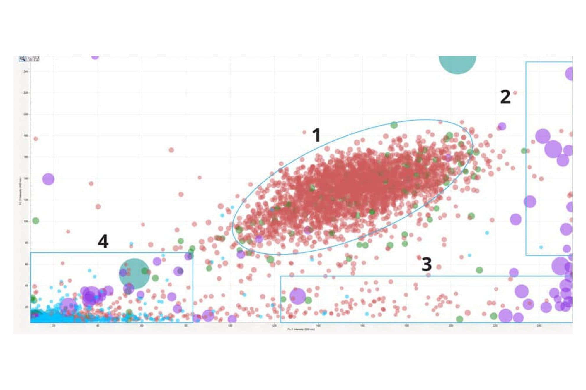
Get fast, definitive particle identification with three fluorescence channels
The Aura+ System comes with three fluorescence channels so you can get the same information from one experiment that would otherwise take three different studies.
In addition, The Aura+ System definitively identifies different types of particles within the same sample for more reliable decision-making:
Three fluorescence membrane microscopy (FMM) channels for three different species of organic particles
Side illumination membrane imaging (SIMI) channel for extrinsic and inorganic particles
Ideal for applications including polysorbate degradation, particulate contamination, protein & antibody workflows, cell therapy workflows, AAV aggregation, AAV stability, cell therapy QC, and cell viability
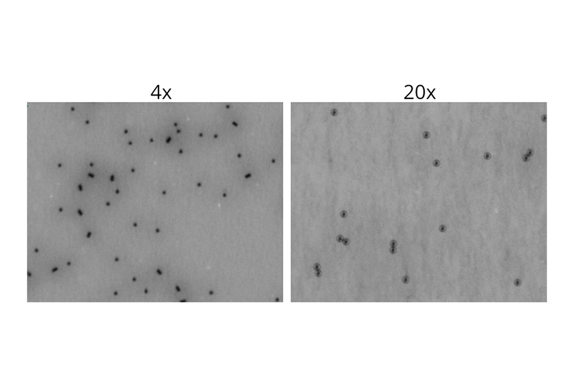
Obtain clear details with high magnification
With the high magnification capabilities of the Aura+ System, you can see more morphological detail of unlabeled particles, enabling decisive discrimination between your lead and undesirable particles.
Quickly differentiate and enumerate cells versus Dynabeads™, quantify polysorbate degradation, and more.
Dynabeads refers to the magnetic beads produced by Thermo Fisher Scientific, Inc. Waters Corporation is not affiliated with Thermo Fisher Scientific, Inc., and references to Dynabeads or any other third-party trademark do not imply sponsorship, endorsement, or approval.
Resources
Documents
What do you want to do?
Frequently Asked Questions
Which method is used to count particles?
Several methods can be used to count particles, depending on factors such as particle size, type, and concentration. Common techniques include microscopy-based methods (such as light microscopy or electron microscopy), flow cytometry, and Coulter counting. The Halo Labs Aura family also provides particle count data, along with size and identification of particles that can impact drug development. Each method has its advantages and limitations, and the choice depends on the specific characteristics of the particles being counted and the analysis requirements.
Why is particle count important?
Particle count is important in various industries, including pharmaceuticals, food, and environmental monitoring, as it provides crucial information about product quality, cleanliness, and safety. In pharmaceuticals, for example, controlling particle count is essential to ensure product efficacy, stability, and compliance with regulatory standards. Similarly, in food processing, monitoring particle count helps ensure product safety and hygiene. In environmental monitoring, particle count can indicate air or water quality and potential health hazards.