Monoclonal antibody (mAb) drug development has been the most active area in the biopharmaceutical industry in recent years. One of the important aspects in recombinant mAb development is to profile glycosylation patterns. Since the glycans play key functions in biological activities, the glycosylation variances during protein production affect the pharmaceutical properties such as efficacy and elimination rate. One of many analytical approaches for glycan analysis is hydrophilic interaction chromatography (HILIC) with fluorescence detection. It provides high sensitivity, good reproducibility, and the ability to separate complex glycan mixtures.
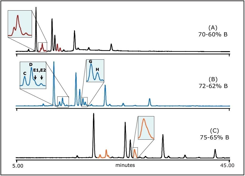
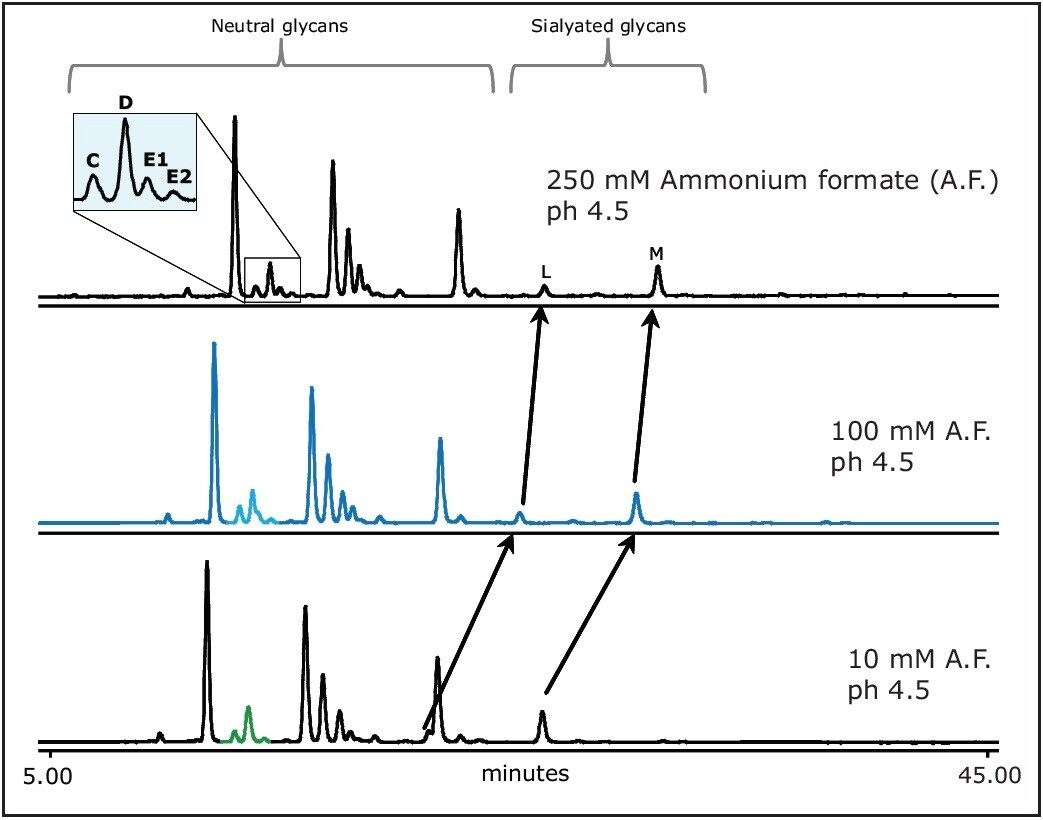
The Waters ACQUITY UPLC System with fluorescence detection (FLR) combined with a Glycan Separation Technology (GST) Column provides superior resolution compared to HPLC systems. These glycan columns, packed with 1.7-μm amide sorbent, efficiently separate the fluorescent-labeled glycans in HILIC mode. Highly-resolved glycan separations, especially for positional isomers and coeluting minor peaks, now can be more accurately measured in UPLC/FLR.
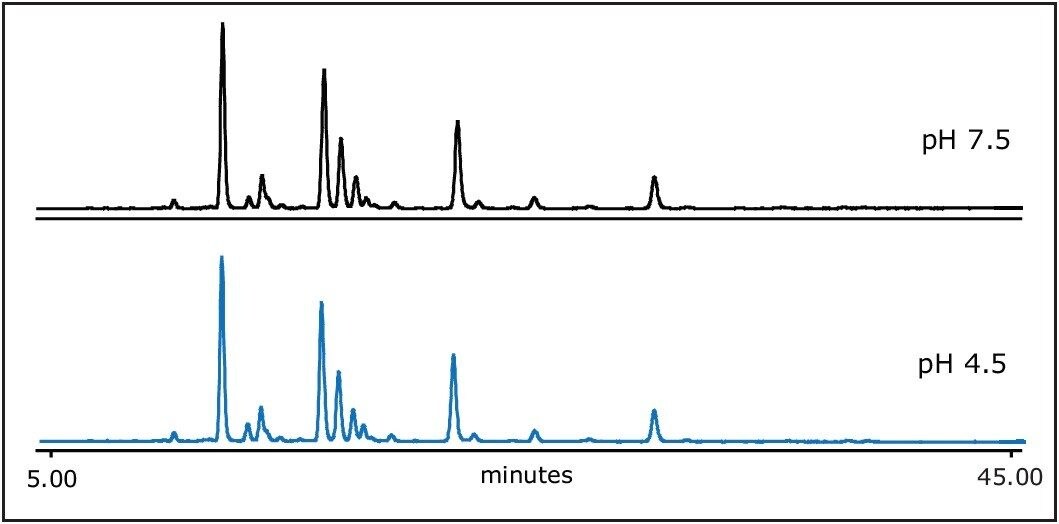
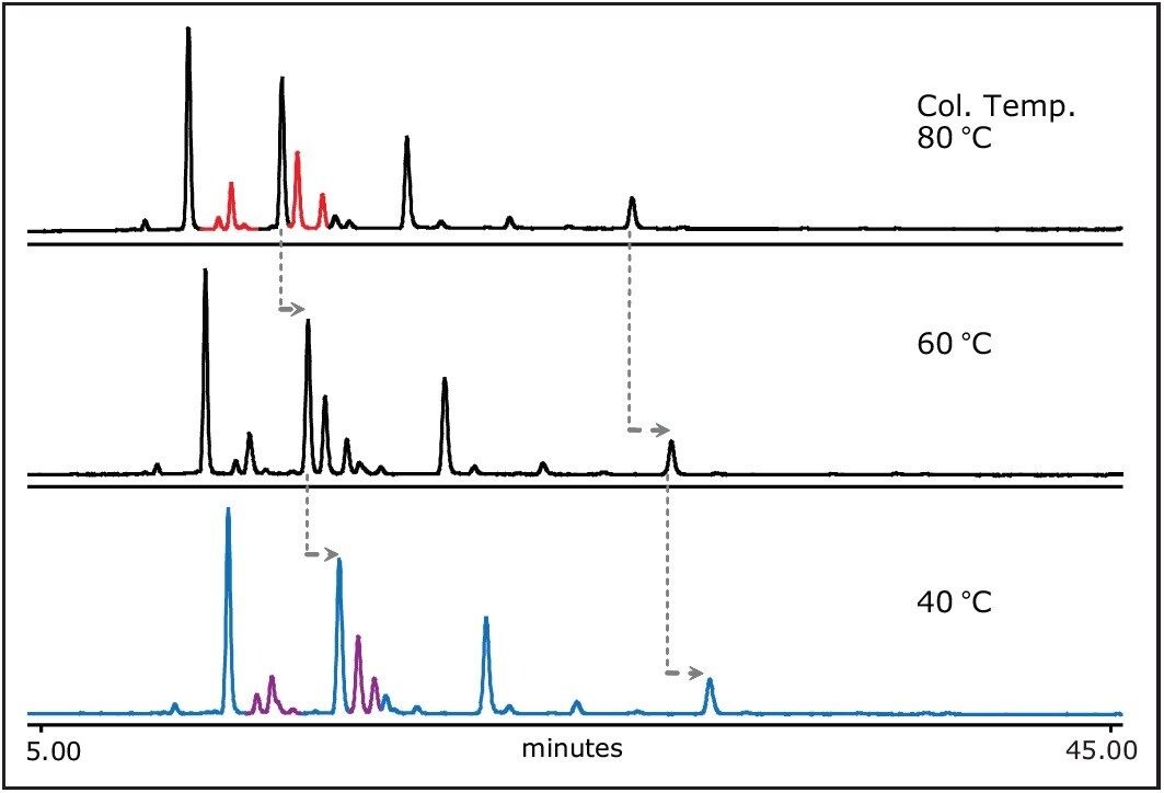
In this application note, a general guideline for researchers to optimize HILIC gradient conditions is shown. The focus is to develop a UPLC method with optimal resolution for 2-aminobenzamide (2-AB) labeled N-linked glycans released from human IgG.
We demonstrate the capabilities of glycan columns to separate fluorescent-labeled glycans in HILIC mode, including the positional isomers and coeluting minor peaks previously unresolved by HPLC.