
Head to Tail Analysis of mRNA by RapiZyme™ MC1 and Cusativin
Alexandre F. Gomes*, Tatiana Johnston*, Balasubrahmanyam Addepalli
* Equal contribution
Waters Corporation, United States
Published on August 10, 2026

Abstract
Continuous innovation in analytical methodologies is essential to address the growing complexity of messenger RNA (mRNA) therapeutic characterization, a field that has expanded rapidly following the success of COVID-19 vaccines. Critical quality attributes (CQAs), including 5′ cap structure and poly(A) tail integrity, play pivotal roles in determining mRNA stability, translational efficiency, and overall therapeutic efficacy. Despite their importance, these attributes are typically assessed using separate analytical workflows that often require multiple enzymatic treatments, extensive sample preparation, and complex data integration. This application note demonstrates the use of the nucleases RapiZyme MC1 and Cusativin to enable a unified digestion strategy capable of simultaneously characterizing multiple key mRNA CQAs — 5′ cap structure, poly(A) tail length, and sequence identity—in a single enzymatic reaction. By consolidating the assessment of these attributes into one streamlined workflow with reduced analytical complexity and improved efficiency, this approach can accelerate comprehensive mRNA quality characterization.
Benefits
- Identification of 5′ capped oligonucleotides and associated impurities
- Assessment of poly(A) tail heterogeneity
- Sequence identity of target mRNA
- Streamlined and speedy workflows for mRNA analysis
- Integrated informatics suite comprising the INTACT Mass, CONFIRM Sequence, Synthetic Library, and MAP Sequence Applications in the waters_connect™ Software environment for CQA analysis
Introduction
Messenger RNA (mRNA)-based therapeutics have advanced rapidly following the success of the Pfizer–BioNTech and Moderna COVID-19 vaccines against SARS-CoV-2. This momentum has accelerated the expansion of RNA medicines beyond infectious diseases, enabling new therapeutic modalities in oncology, rare genetic disorders, and cell engineering.1 Emerging applications include personalized anticancer mRNA vaccines; genome-editing strategies for conditions such as sickle cell disease; protein-replacement approaches for metabolic disorders like propionic acidemia; in vivo production of therapeutic antibodies; and mRNA-engineered CAR-T cell therapies.
Therapeutic mRNA is produced by in vitro transcription (IVT), a process that demands rigorous characterization of CQAs, including purity, structural integrity, sequence identity, safety, and functional potency.2 Integrity of the protein coding open reading frame (ORF) is critical for the successful production of target protein by mRNA therapeutics. Efficient translation of ORF is highly dependent on the presence of a correctly formed 5′ cap structure because of its interactions with ribosome. The 5′ cap may be incorporated co-transcriptionally or introduced enzymatically post-transcription, and its structural integrity plays a central role in governing mRNA stability, translational efficiency, and overall protein expression.
In addition to the 5′ cap, the poly(A) tail is a critical structural element that influences the stability and translational efficiency of therapeutic mRNA. The poly(A) sequence can be encoded directly within the DNA template — whether plasmid-based or PCR-derived — or appended enzymatically in a separate post-transcriptional step.3 Regardless of the production method, an appropriately sized and less heterogeneous poly(A) tail is essential for maintaining mRNA stability, promoting efficient ribosome recruitment, and enabling sustained protein expression following intracellular delivery.3
Variability in cap structure, incomplete capping, heterogeneity in poly(A) tail length and composition, and by-products arising from capping or tailing reactions can significantly impact mRNA stability, translation efficiency, and overall therapeutic potency.4 As mRNA applications continue to expand and regulatory expectations become more stringent, there is an increasing need for robust, sensitive, and high-resolution analytical methods capable of comprehensively characterizing both 5′ cap quality and poly(A) tail integrity apart from sequence identity. Addressing these challenges is critical to ensuring consistent mRNA performance and advancing the development of next-generation RNA therapeutics.
This application note demonstrates that the ribonucleases RapiZyme MC1 and Cusativin enable a unified digestion workflow that resolves key mRNA CQAs, —including 5′ cap structure, poly(A) tail heterogeneity, and sequence identity, offering a streamlined and efficient approach to mRNA characterization.
Experimental
Materials
N,N-Diisopropyethylamine (DIPEA, 99% purity, catalog# D214752-500ml, Millipore Sigma, St. Louis, MO) and 1,1,1,3,3,3-hexafluoro-2-propanol (IonHance™ HFIP, Waters, p/n: 186011465) were used. HPLC grade type I deionized (DI) water was purified using a Milli-Q™ System (Millipore, Bedford, MA). Mobile phases were always prepared fresh (same day) just before the data acquisition. Ultrapure nuclease-free water (Thermo Fisher Scientific (Waltham, MA) catalog# J71786.AE) was used for mRNA digestion.
mRNA Digestion
Firefly-Luciferase (FLuc) mRNA (Cap1, m1Ψ) catalog# RP-A00023-0.2 and its unmodified version (custom order) were purchased from GenScript (Piscataway, NJ).
For mRNA digestion with RapiZyme RNase MC1 (Waters, p/n: 186011190) or Cusativin (Waters, p/n: 186011192), 20 µg of mRNA was denatured by heating at 90 ºC for 2 minutes and quickly cooled on ice for 2 minutes. Approximately 300 U of enzyme for unmodified RNA (15 U/µg) and 600 U (30 U/µg) for m1Ψ modified mRNA were then added, with a total digestion volume of 60 µL, in a buffered 100 mM ammonium acetate diluent with either pH 8.0 for MC1 or pH 9.0 for Cusativin. After incubation of the digestion mix at 30 ºC for 60 minutes, the enzymes were heat inactivated at 70 ºC (MC1) or 75 ºC (Cusativin) for 15 minutes. The resulting reactions were then transferred to sample vials and analyzed by LC-MS. For more detailed information on RapiZyme MC1 and RapiZyme Cusativin, see accompanying care and use manual and application notes.6
Ion Pairing Liquid Chromatography (IP-RP-LC) Conditions
|
LC-MS system: |
Xevo™ MRT with ACQUITY™ Premier UPLC™ System (Binary Solvent Manager, Sample Manager FTN, ACQUITY TUV Detector) |
|
Column: |
ACQUITY Premier Oligonucleotide BEH™ C18 Column, 130 Å, 1.7 µm, 2.1 mm x 150 mm (Waters p/n: 186009486) |
|
Column temperature: |
70 °C |
|
Flow rate: |
0.4 mL/min |
|
Mobile phases: |
Mobile phase A: 0.1% DIPEA (N,N-Diisopropylethylamine), 1% HFIP (1,1,1,3,3,3-hexafluoroisopropanol) in water Mobile phase B: 0.0375% DIPEA, 0.75% HFIP in water/acetonitrile 35:65 |
|
Sample temperature: |
8 °C |
|
Sample vials: |
QuanRecovery™ Vials with MaxPeak™ High Performance Surfaces (HPS) (Waters, p/n: 186009186) |
|
Injection volume: |
10 µL |
|
Wash solvents: |
Purge solvent, Sample Manager wash solvent, Seal wash: 20% acetonitrile in DI water |
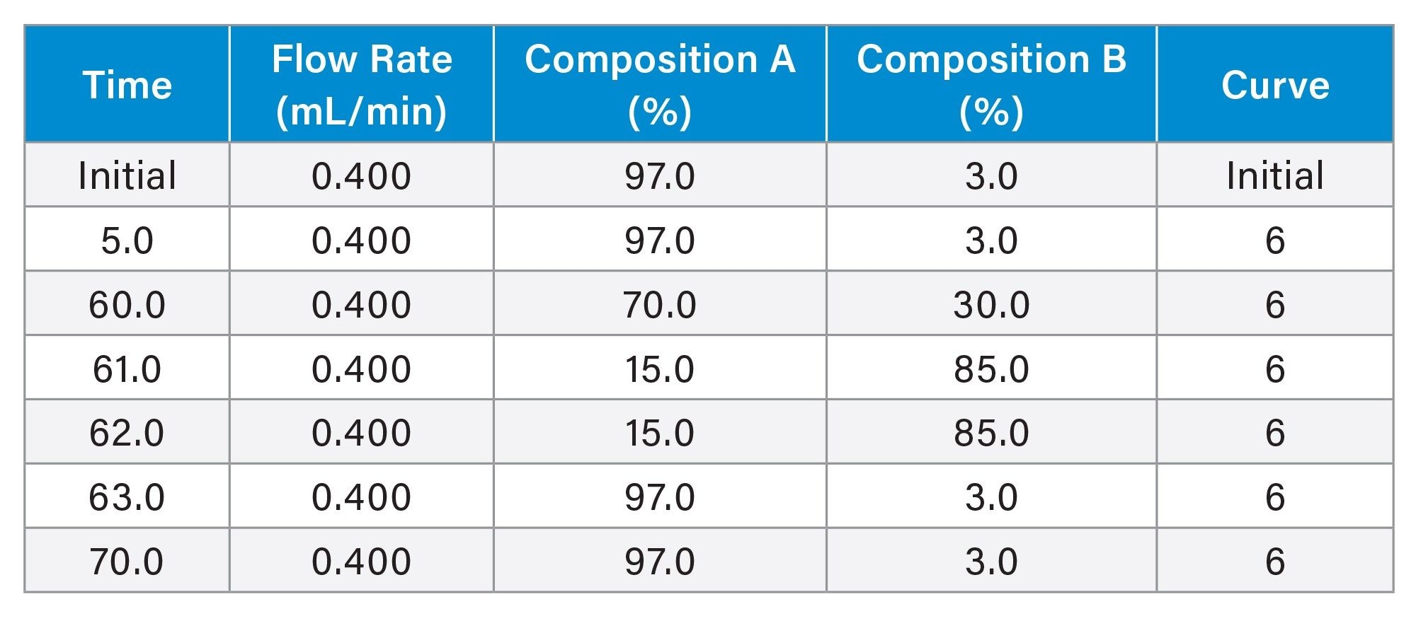
Gradient Table
MS Conditions
|
Mass spectrometer: |
Xevo MRT Mass Spectrometer (176005403) |
|
Ionization mode: |
ESI- |
|
Capillary voltage: |
1.5 kV |
|
Cone voltage |
40 V |
|
Source temperature: |
120 °C |
|
Desolvation gas flow: |
1000 L/h |
|
Desolvation gas temperature: |
550 °C |
|
Quad profile: |
600/700/800 (20:20:20:40) |
|
Collision cell RF: |
1200 V |
|
Acquisition mode: |
MSE (DIA) |
|
Acquisition range: |
m/z 50 - 4000 |
|
Scan time: |
0.5 s |
|
Low collision energy: |
6 V |
|
High collision energy ramp: |
25 - 45 V |
|
IDC: |
Low (5) |
|
Lock correction: |
Automatic (Single Point) |
|
Data analysis: |
waters_connect Software v.4.0, INTACT Mass App v.1.9, Synthetic Library v. 2.0, CONFIRM v.2.0.0 and MAP Sequence v.2.0 |
Data Processing
All data processing was performed using waters_connect Software v.4.0.0. LC-MS data sets were used for 5’ capping, sequence integrity and poly(A) tail analysis.
The 5′ capping analysis of modified FLuc (Cap1, m1Ψ) and unmodified FLuc (Cap1, custom-made) sample sets were processed in the INTACT Mass Application using an analysis method configured to identify the expected Cap1 structure as well as potential in vitro transcription (IVT)-derived 5′ cap impurities. Structural annotations of capping products were subsequently verified with MS/MS fragment ion data using the CONFIRM Sequence Application.
For the sequence integrity analysis (deciphering sequence), in silico digestion of the mRNA sequence with RapiZyme MC1 and Cusativin was performed in Synthetic Library to support the experimental design. Digestion products generated by both enzymes were then confirmed in MAP Sequence, and combined sequence coverage was assessed using the Exclude Ambiguities option to provide overall sequence coverage metrics.
For the poly(A) tail analysis, digests generated with both RapiZyme MC1 and Cusativin were evaluated. Poly(A) tail masses were successfully deconvoluted, and heterogeneity profiles were determined for both unmodified and modified FLuc mRNA samples using either enzyme. However, Cusativin is generally the preferred enzyme for poly(A) tail characterization due to its broad but more suitable cleavage specificity. Poly(A) tail analysis was performed using the INTACT Mass Application.
Results and Discussion
Analysis of MC1 Digests Reveals the Presence of 5′ Cap1-related Digestion Products and IVT Capping Impurities
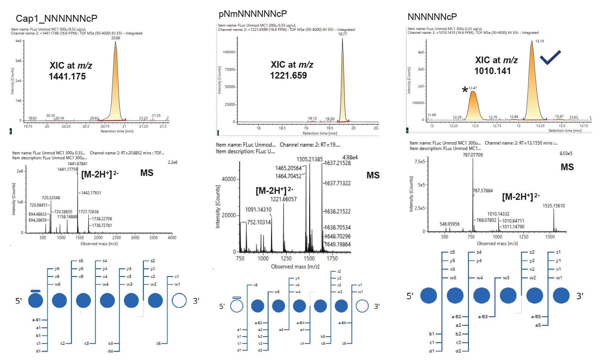
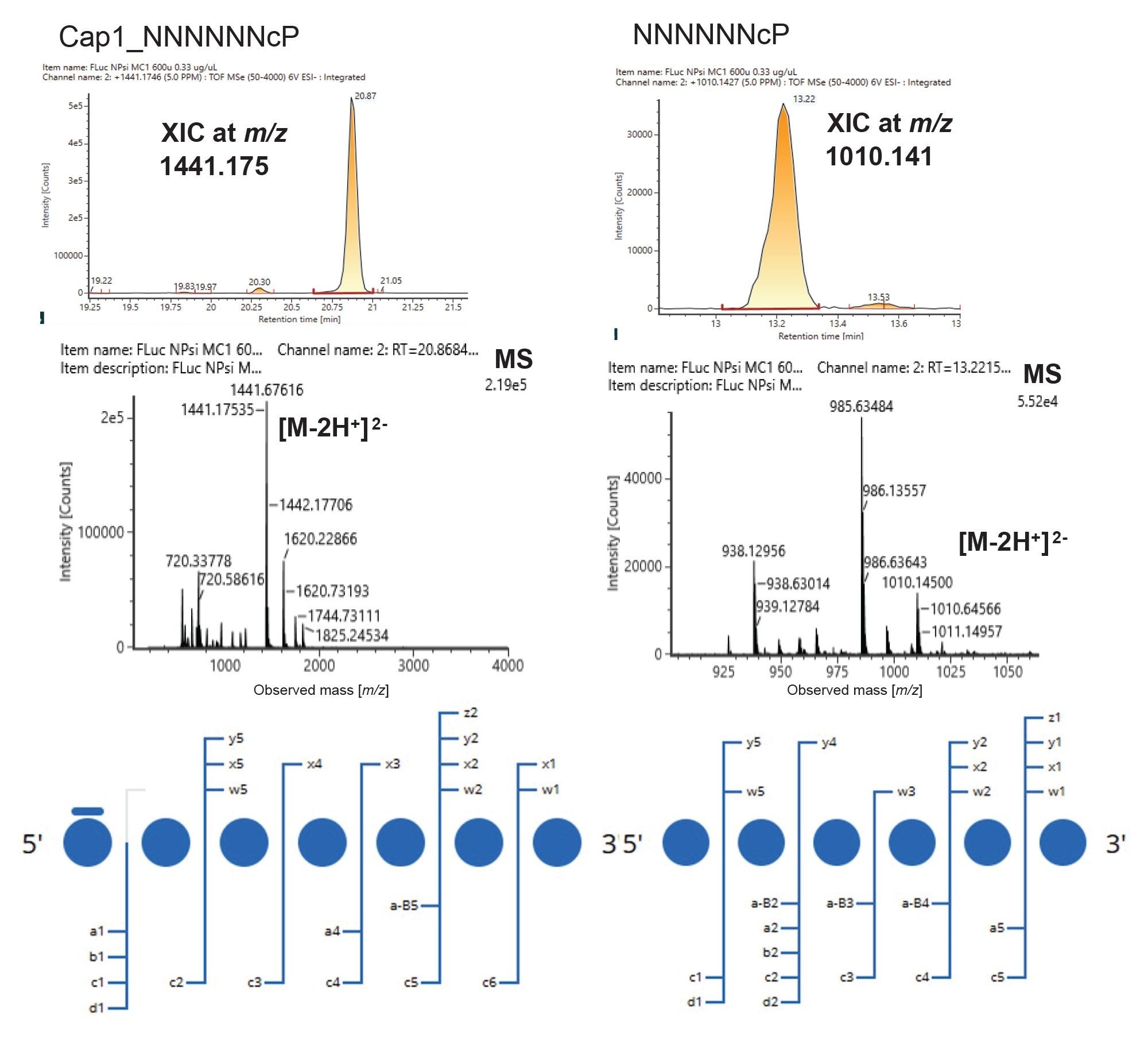
The 5′ UTR sequence of the FLuc mRNA is proprietary and is therefore not disclosed in this report. However, based on the digestion rules of the enzymes, a digestion product with six nucleotides apart from the cap is expected in the cap-containing or no cap oligonucleotides for both versions of unmodified FLuc and m1Ψ-modified mRNA constructs. The digestion product with fully capped species was detected as a [M−2H]²⁻ ion at m/z 1441.175 and was assigned to Cap1_NNNNNNcP (Figure 1). This species eluted at RT 20.89 minutes for the unmodified FLuc mRNA (Figure 1) and at RT 20.87 minutes for the m1Ψ-modified FLuc mRNA (Figure 2). A corresponding uncapped species was also detected in both unmodified and modified FLuc mRNA samples, eluting at RT 13.16 minutes and 13.22 minutes with the same m/z value (1010.141) and charge state ([M−2H]²⁻). In addition, a pNm_NNNNNNcP capping-related impurity was detected exclusively in the unmodified FLuc mRNA sample. This species eluted at RT 19.77 minutes and was observed as a [M−2H]²⁻ ion at m/z 1221.659 (Figure 1). The assignments are further corroborated by the scoring of product ions for each nucleotide position in the sequence as shown in the bottom panels.
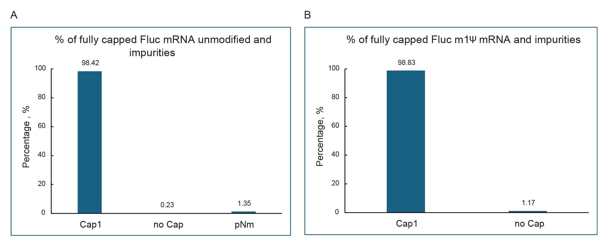
Capping efficiency was determined from the extracted ion chromatogram (XIC) peak areas of capped and uncapped digestion products, along with any additional detected impurities, using waters_connect Software. Analysis of 5′ capping and associated impurities using RapiZyme MC1 digestion profiles identified Cap1 (98.42%), pNm (1.35%), and uncapped mRNA (0.23%) versions in unmodified FLuc mRNA, and Cap1 (98.83%) and uncapped mRNA (1.17%) versions in m1Ψ-modified FLuc mRNA. The low levels of capping-related impurities indicate high capping efficiency for both constructs (Figure 3).
mRNA Sequence Identity Confirmation by RapiZyme MC1 and RapiZyme Cusativin Through LC-MS
For sequence identity assessment, Synthetic Library was used to predict sequence coverage from in silico digestion products generated for FLuc mRNA using RapiZyme MC1 and Cusativin as the digestion enzymes. After importing the FLuc mRNA sequence, processing parameters were configured according to Waters Application Note, 720009171.9 Sequence coverage was evaluated using digestion products from both enzymes to maximize coverage in a single workflow.
MAP Sequence utilizes both MS1 precursor and MS2 fragment ion data acquired by MSE to assign digestion products, calculate unique sequence coverage, and identify potential alternative assignments or ambiguities. The poly(A) tail was excluded from MAP Sequence processing and peak assignment; instead, INTACT Mass App analysis was used to characterize poly(A) tail length and heterogeneity.
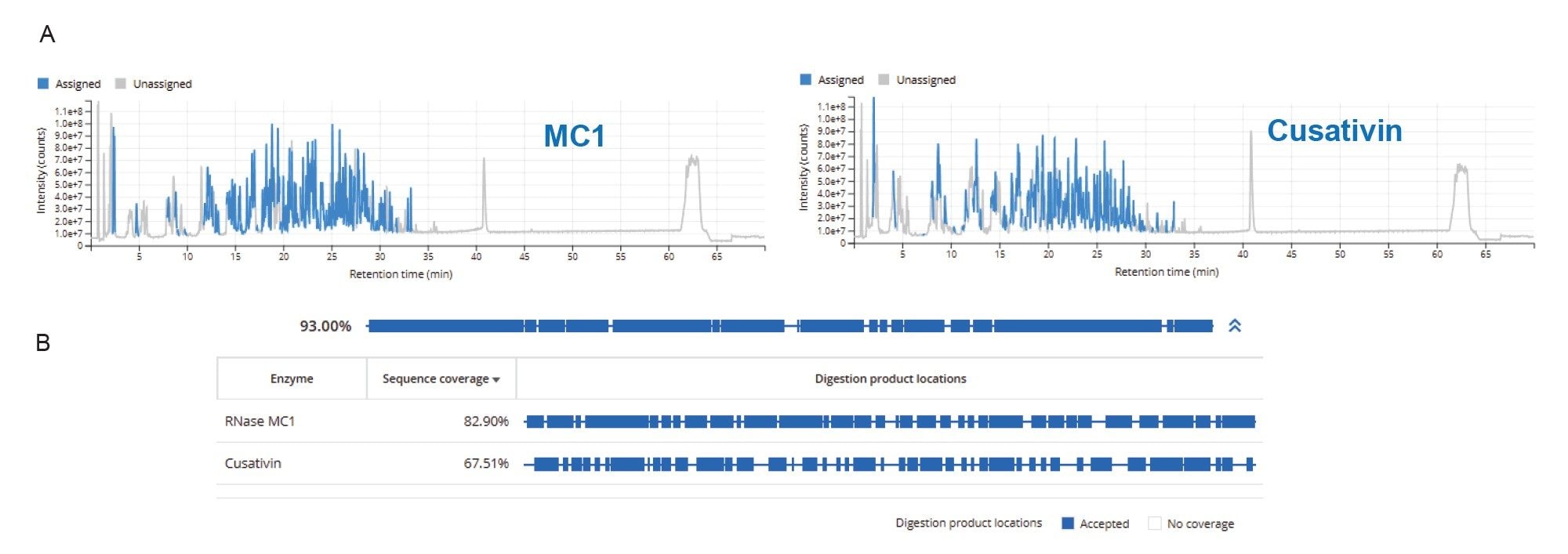
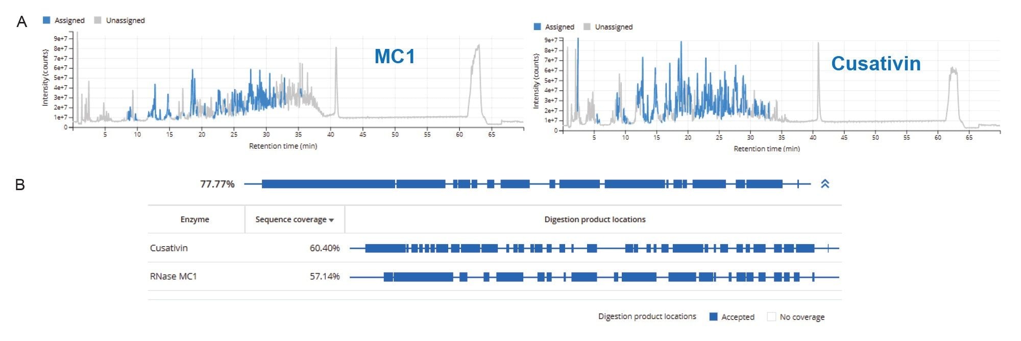
Using this approach, a combined MC1/Cusativin sequence coverage of 93% (excluding cap and poly(A) tail) was achieved for unmodified FLuc mRNA under the digestion and LC-MS conditions evaluated. Individually, MC1 and Cusativin provided sequence coverages of 82.9% and 67.5%, respectively (Figure 4). MAP Sequence enabled the integration of complementary information from both digests into a single coverage map, substantially increasing overall sequence coverage. For m1Ψ-modified FLuc mRNA, the combined MC1/Cusativin sequence coverage was 77.8% (Figure 5). Identified oligonucleotide products are highlighted in the TIC trace for both enzymes. Interestingly, there are a few peak traces that are not assigned in both MC1 and Cusativin digests and manual verification of these peaks could potentially improve the sequence coverage.
These results demonstrate that combining sequence coverage data from complementary MC1 and Cusativin digestions can provide a more complete mRNA sequence characterization and is particularly advantageous for the analysis of longer RNA molecules.
Poly(A) Tail Length and Heterogeneity Analysis
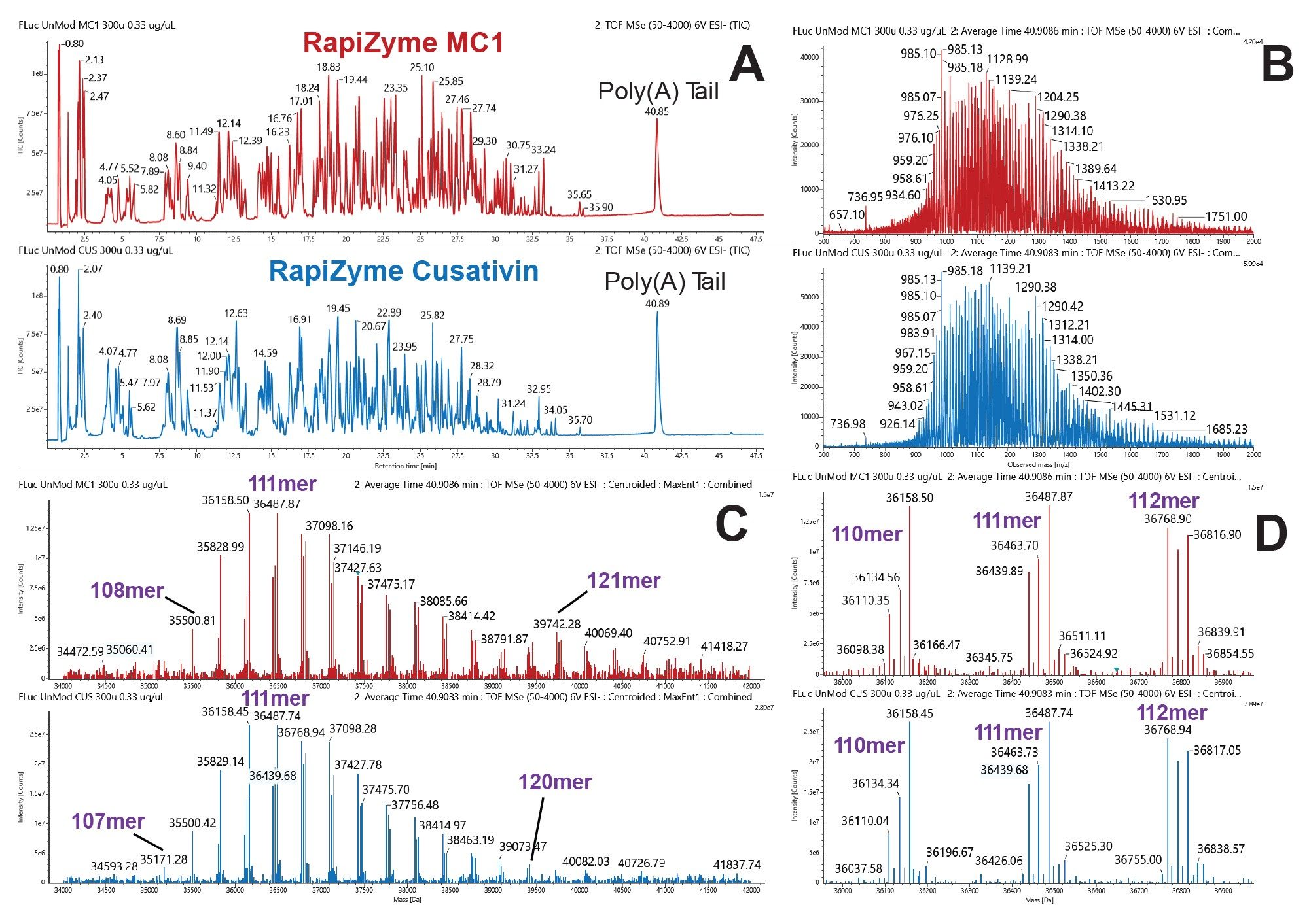
For both unmodified and m1Ψ-modified FLuc mRNAs, a distinct poly(A) tail peak was observed at RT 40.85–40.89 minutes. Although the raw mass spectra were complex, deconvolution with MaxEnt1 (waters_connect Software) resolved a distribution of species between 35 and 41 kDa. These masses were consistent with a 5′ GGC sequence followed by a poly(A) tail ranging from 104–114 nt, centered on a 111-mer (36,487 Da) (Figure 6). Further processing in INTACT Mass App enabled assignment of individual poly(A) tail lengths and determination of their relative abundances.
The poly(A) tail spectra also revealed sequence heterogeneity. Examination of the deconvolved spectra showed that each tail length was represented by a triplet of peaks with mass shifts of −24 Da and −48 Da, consistent with the presence of one or two rA→rC substitutions, respectively. These results indicate that the FLuc mRNA poly(A) tails comprise a mixture of homogeneous poly(A) species and variants containing one or two cytidine substitutions. Such heterogeneity is consistent with the known ability of E. coli poly(A) polymerase (PAP) to incorporate CTP, and to a lesser extent UTP, during polyadenylation.9 In addition, LC–MS analysis enabled detection and confirmation of these poly(A) tail sequence variants, demonstrating the utility of this approach for characterizing poly(A) tail composition and heterogeneity.
Conclusion
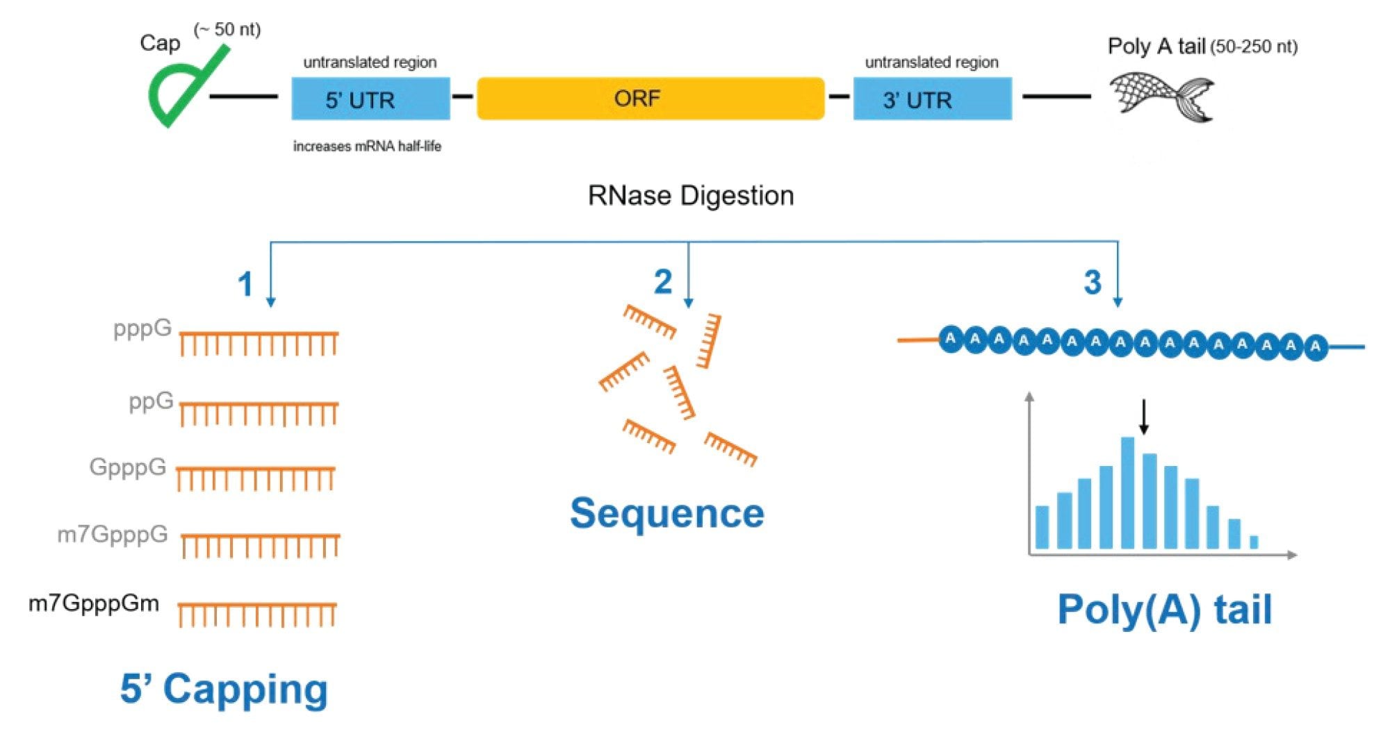
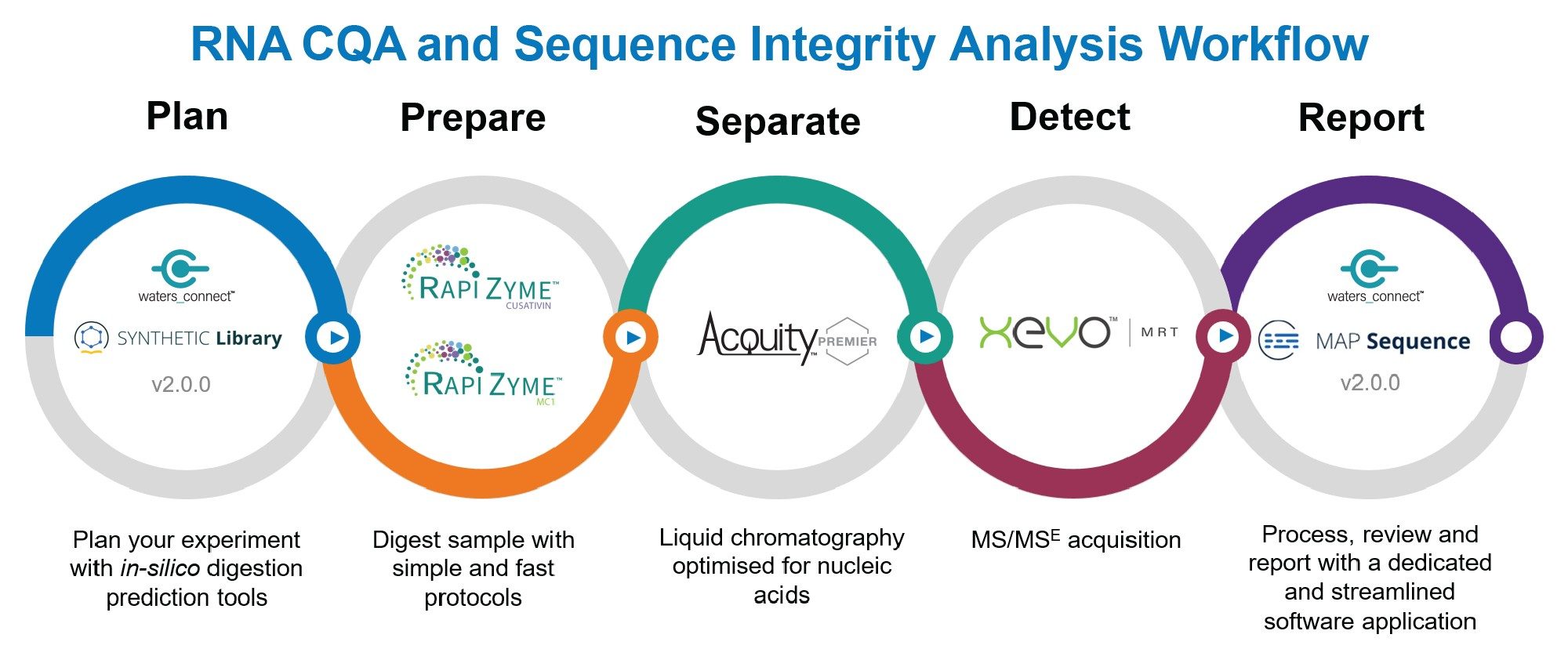
In this work, the utility of RapiZyme MC1 and RapiZyme Cusativin for comprehensive mRNA CQA analysis are demonstrated, including 5′ capping efficiency, poly(A) tail length and heterogeneity, and sequence identity, using a streamlined digestion workflow (Figure 7) coupled with high-resolution Xevo MRT Mass Spectrometry and Waters informatics tools (CONFIRM, INTACT Mass App, Synthetic Library, and MAP Sequence). This integrated approach enables comprehensive mRNA characterization through complementary cleavage specificities and sequence coverage.
References
- Qin, S. et al. mRNA-based therapeutics: powerful and versatile tools to combat diseases. Signal Transduct. Target. Ther. 2022, 7, 166.,
- Camperi, J. et al. Comprehensive impurity profiling of mRNA: evaluating current technologies and advanced analytical techniques. Anal. Chem. 2024, 96, 3886-3897
- Stepinski, J. et al. Synthesis and properties of mRNAs containing the novel “anti-reverse” cap analogues 7-methyl(3”-O-methyl) GpppG and 7-methyl(3’-deoxy) GpppG. RNA 2001, 7,1486-1495
- Trepotec, Z. et al. Segmented poly (A) tails significantly reduce recombination of plasmid DNA without affecting mRNA translation efficiency or half-life. RNA 2023, 25, 507-518
- Fuchs, A. L. et al. A general method for rapid and cost-efficient large-scale production of 5’ capped RNA. RNA 2016, 22, 1454-1466
- Addepalli, B. et al. Tunable digestions of RNA using RapiZymes RNases to confirm sequence and map modifications. Waters application note, 720008539, Sep 2024
- Addepalli, B. et al. Analysis of mRNA cap impurities profiles and capping efficiency using RapiZyme MC1 ribonuclease. Waters application note, 720008793, June 2025
- Doneanu, C. et al. Sequence mapping of mRNA digests using Xevo™ MRT Mass spectrometer and waters_connect™ MAP Sequence 2.0 application. Waters application note, 720009171, Dec 2025
- Yehudai-Resheff, S; Schuster, G. Characterization of the E. coli poly(A) polymerase: nucleotide specificity, RNA-binding affinities and RNA structure dependence. Nucleic Acids Res. 2000, 28(5), 1139-1144
Featured Products
720009517, August 2026


