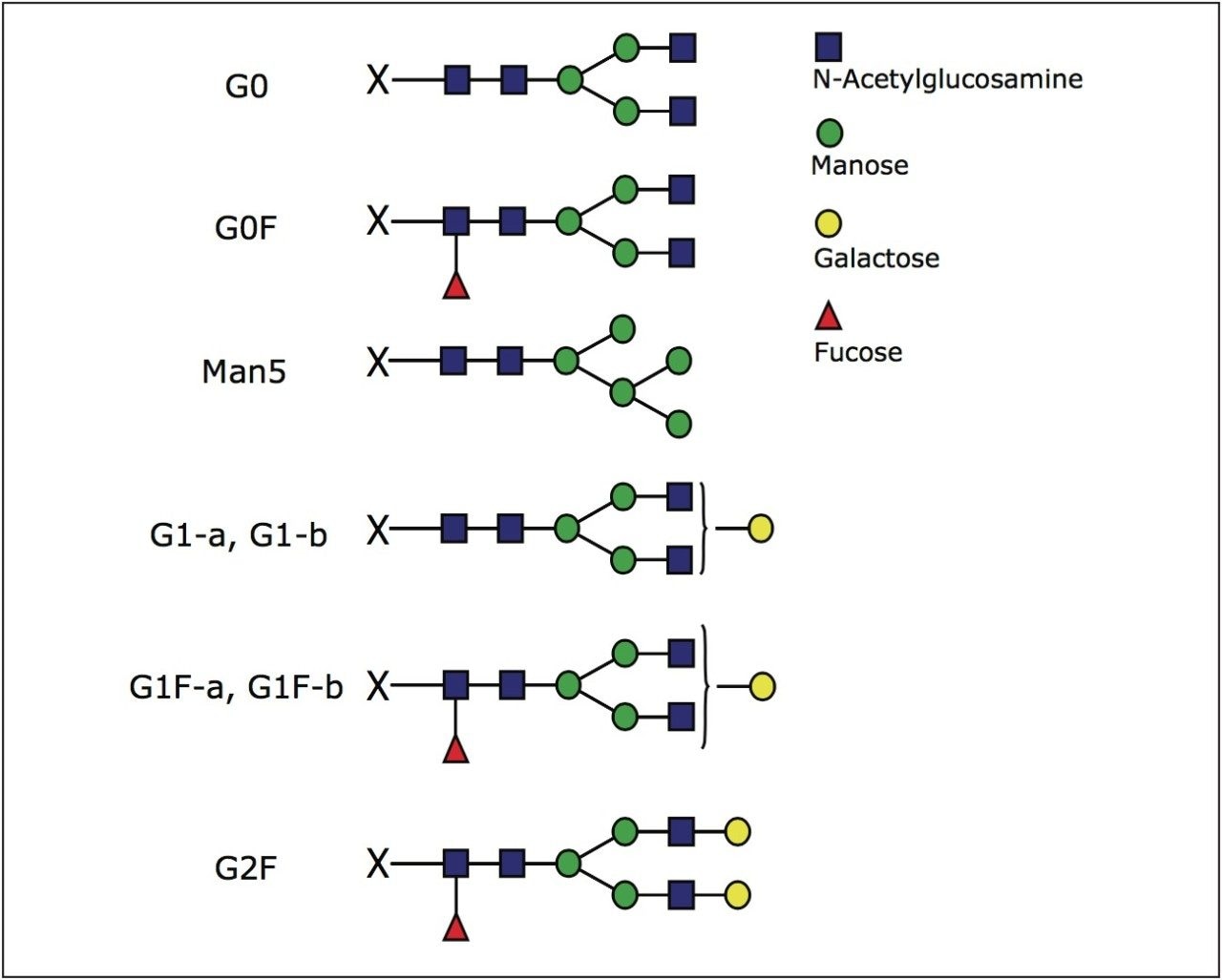
Glycosylation of proteins affects their tertiary structure and potentially therapeutic efficacy. Therefore, the glycosylation of therapeutic proteins such as monoclonal antibodies (mAb) needs to be closely monitored.
Reversed-phase liquid chromatography (RP-LC) is a primary method chosen for protein characterization via peptide mapping. Peptide mapping applications require efficient columns to resolve complex peptide mixtures into unique peptides. Modified peptides, such as oxidized or deamidated ones, can also be separated from the unmodified peptides.1 UltraPerformance Liquid Chromatography (UPLC) technology provides the resolving power needed for these challenging separations.2
It has been reported that RP-UPLC is able to resolve glycosylated peptides into their glycoforms.3 However, the complete resolution of glycopeptide micro-heterogeneity (same peptide sequence, various glycoforms) remains difficult. This is because retention in RP-LC is mainly due to peptide hydrophobicity, and is less affected by the presence of hydrophilic glycans. The separation is further complicated by the presence of non-glycosylated peptides in the sample that often elute in the vicinity of the glycopeptides of interest.
Several separation methods are available for glycan analysis, including capillary electrophoresis (CE), high pH anion exchange chromatography with pulsed amperometric detection (HPAEC-PAD), and hydrophilic interaction chromatography with fluorescent detection of labeled glycans (HILIC/FLR). While those methods are useful, the confirmation of glycan identity relies on their retention time, available standards, and use of specific exoglycosidase enzymes.4 Fraction collection of resolved glycans is often combined with a matrix assisted laser desorption/ionization (MALDI) MS method for confirmation of mass of glycans and their MS/MS structure identification. Because of the advantages of on-line MS, the off-line MALDI method is being recently replaced with LC-MS glycan analysis.
Two LC-MS methods currently under development are MS analysis of the intact proteins and LC/FLR-MS analysis of the glycan released from a glycoprotein. In the first case, the mass spectrum (after deconvolution) provides information about the protein molecular weight and its heterogeneity due to glycosylation.5 For mAbs, where the glycosylation nature is well understood, the intact mass information can be translated into the relative quantitation of glycoforms.6 Though useful as fast screening, the intact protein MS method may fail to detect minor glycoforms.
The second method for glycoprotein characterization utilizes specific enzymes (PNGase F) to release N-linked glycans from the protein. Glycans are typically enriched, labeled with fluorescent dye, and analyzed in HILIC mode. Highly efficient UPLC HILIC columns have been shown to facilitate an excellent glycan separation and relative quantification.7
HILIC separation of glycans is considered to be a reliable method. However, for proteins with multiple N-linked glycosylation sites, released glycans of the same type elute in chromatogram as cumulative peaks. Therefore, the information about the occupancy of different N-linked sites is lost. This is also the case for CE and HPAEC-PAD methods. While this does not present a problem for proteins with a single glycosylation site, such as monoclonal antibodies, it precludes full characterization of proteins with multiple glycosylation sites.
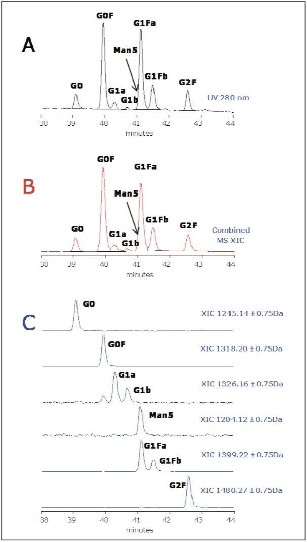
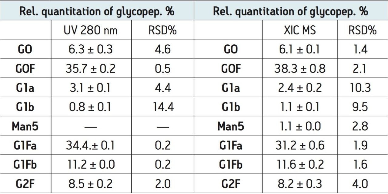
In this application note we propose an orthogonal method, UPLC HILIC/TUV-MS, in which the information about glycan heterogeneity and site occupancy is preserved. This method is complementary to UPLC HILIC/FLR analysis of the released glycans and the RP peptide map. The same tryptic digest used for the peptide map can be used in the method. The ACQUITY UPLC System with a UPLC BEH Glycan Column is used for UPLC/FLR analysis of released glycans and the proposed method7 for the separation of glycopeptides.3