
Detect Specific Protein Aggregates in Biologic Formulations with the Aura Immunoassay
Published on October 07, 2026

Introduction
Antibodies are widely used in immunoassays to reliably target and identify a particular protein of interest. Proteins are labeled with a primary antibody specific to the protein and detected with a secondary antibody conjugated to a chemiluminescent or fluorescent molecule. Countless applications using antibody-protein interactions have been developed for pharmacokinetic/pharmacodynamic (PK/PD) assays to identify and quantify protein content. The Aura™ aggregate analysis system utilizes fluorescence membrane microscopy (FMM) to identify particles stained with a fluorescent dye. For example, proteinaceous particles stained with Thioflavin T (ThT) can be differentiated from other non-proteins, as described in Application Note 7, Rapidly Distinguish Protein from Non-Protein Particles in Biologic Formulations. Labeling protein particles with fluorescently tagged antibodies enable specific and multiplexed protein identification of sources of stability issues in co-formulated antibody therapeutics in conjunction with subvisible particle information.
This application note introduces a two-step fluorescent antibody labeling method that identifies aggregates of a specific protein in a mixed sample with a high degree of specificity and accuracy on the Aura System. The method is robust and can be used with proteins labeled both in their native liquid state as well as post filtration.
Experimental
Sample Preparation
Human IgG (HIgG) and hen egg-white lysozyme (HEWL) proteins were antibody labeled using a solution phase or membrane phase protocol before imaging on the Aura System. In both methods, ThT was applied to each well after the initial image acquisition to determine the total protein content to corroborate the antibody labeling efficiency and accuracy. ThT staining and IgG labeling were imaged using the FL1 and FL2 channel, respectively.
Solution Phase Labeling
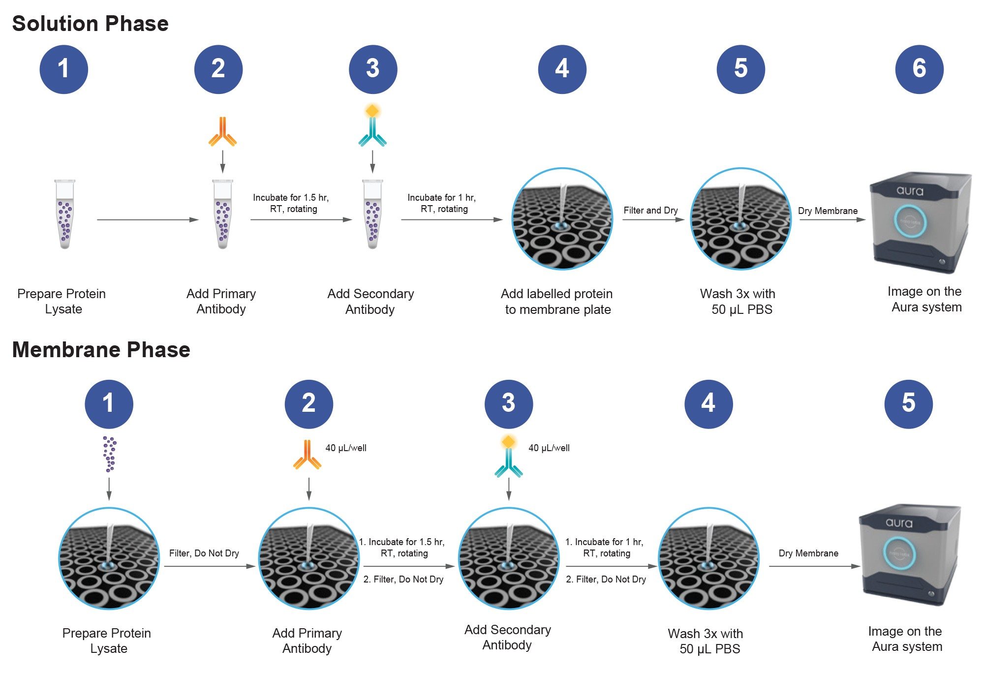
50 µg/mL HIgG or HEWL were each incubated in a 1.5 mL Eppendorf tube with rabbit monoclonal recombinant Human-IgG antibody (AbCam ab109489) diluted 1:200 in PBS for 90 minutes at room temperature with rotation (Figure 1). After the primary incubation, Alexa Fluor® 488 conjugated goat anti-rabbit IgG (AbCam ab150077) diluted 1:100 in PBS was added to the solution and incubated at room temperature for 60 minutes with rotation. An antibody only sample was used as a negative control. 35 µL of each sample was applied to a black membrane plate well and measured in triplicate. For the mixed HIgG + HEWL sample, 35 µL of labeled HIgG was first filtered through the membrane before 35 µL of labeled HEWL was applied. All samples were washed 3x with PBS to reduce non-specific binding prior to imaging. Bovine IgG (BIgG) was used as a separate control for multiplexed solution phase labeling to assess co-formulation application feasibility.
Membrane Phase Labeling
35 µL of HIgG, HEWL, or 35 µL of HIgG + HEWL was applied to corresponding wells in a black membrane plate in triplicate. Antibody only was used as a negative control. 50 µL of the HIgG antibody (1:200 dilution in PBS), were applied to each well and left to sit but not vacuumed for 90 minutes. Then, 50 µL of secondary antibody (1:100 in PBS) was applied to the membrane for 60 minutes before filtering the plate. All sample wells were washed with PBS prior to imaging.
Results and Discussion
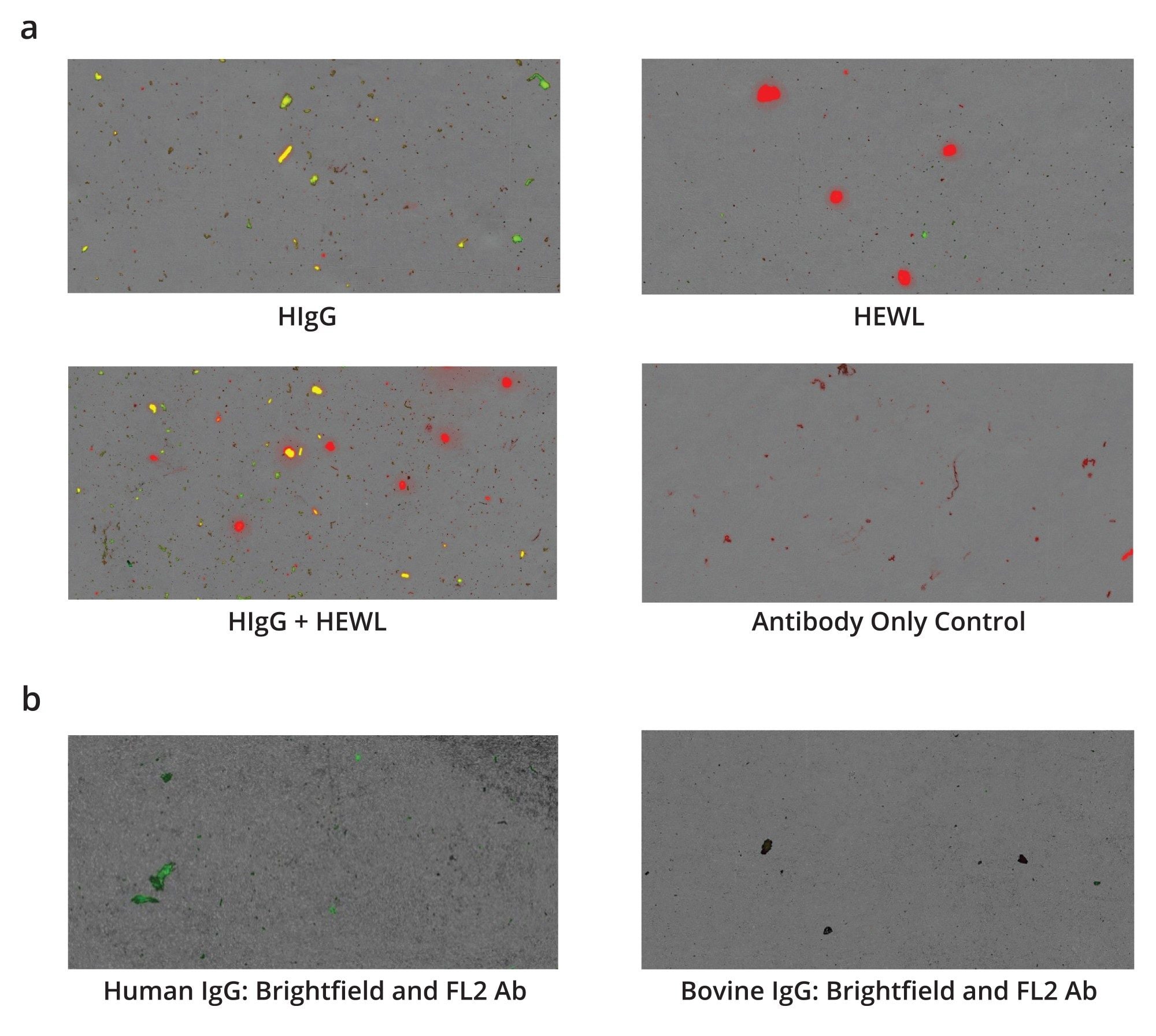
Samples that were antibody labeled using the solution phase protocol were imaged on the Aura System (Figure 2a). The red signal in the combined brightfield and fluorescent image represents total protein content stain by ThT, green designates proteins fluorescently labeled with antibody, and the yellow represents the combination of both signals.
Most of the signal in the HIgG well (top left) is yellow indicating these are HIgG protein particulates. In the HEWL image (top right), the protein is only stained red with ThT (total protein) revealing that while there is protein, it is not HIgG. The HIgG + HEWL mixed sample (bottom left) shows the ability of the Aura System to specifically recognize HIgG protein aggregates in a solution containing HIgG (yellow) and HEWL (red). While the primary antibody only control (bottom right) does form some protein aggregates, there is no discernable fluorescent signal due to secondary antibody labeling.
To demonstrate feasibility for multiple antibody co-formulation applications, samples were labeled with HIgG and Bovine IgG primary antibody. The assay discerns between two different antibodies and isn’t limited to just differentiating between two different proteins (Figure 2b). Human IgG on the left displays significant staining in the FL2 channel, while Bovine IgG does not.
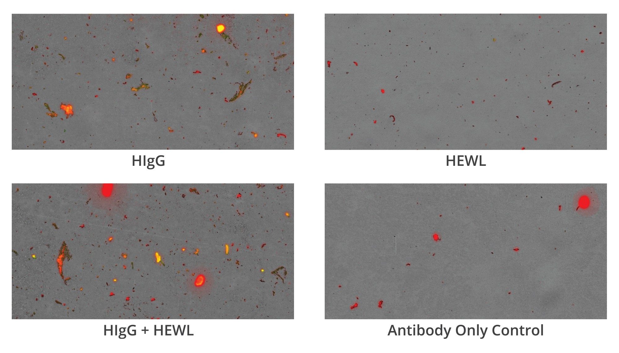
Antibody samples labeled using the membrane phase labeling protocol were also imaged on the Aura System (Figure 3). Again, the red signal in the fluorescent images represents total protein stain by ThT, green is antibody fluorescence, and yellow is the combination of both signals. Most of the signal in the HIgG well (top left) is yellow indicating that these are HIgG protein particulates, just like in solution phase stained samples. In the HEWL image (top right), the lysozyme protein is only stained red with ThT (total protein) suggesting that while it is a protein, it is not antibody labeled HIgG. The HIgG + HEWL combination (bottom left) shows how Aura can differentiate between different proteins in a lysate containing multiple proteins, as HIgG protein is dually labeled and stained with antibody and ThT, respectively (yellow) and the HEWL is only stained with ThT (red). While the antibody only control (bottom right) does form some protein aggregates, there is no discernable fluorescent signal due to secondary antibody labeling.
Quantitative Analysis and Assay Accuracy
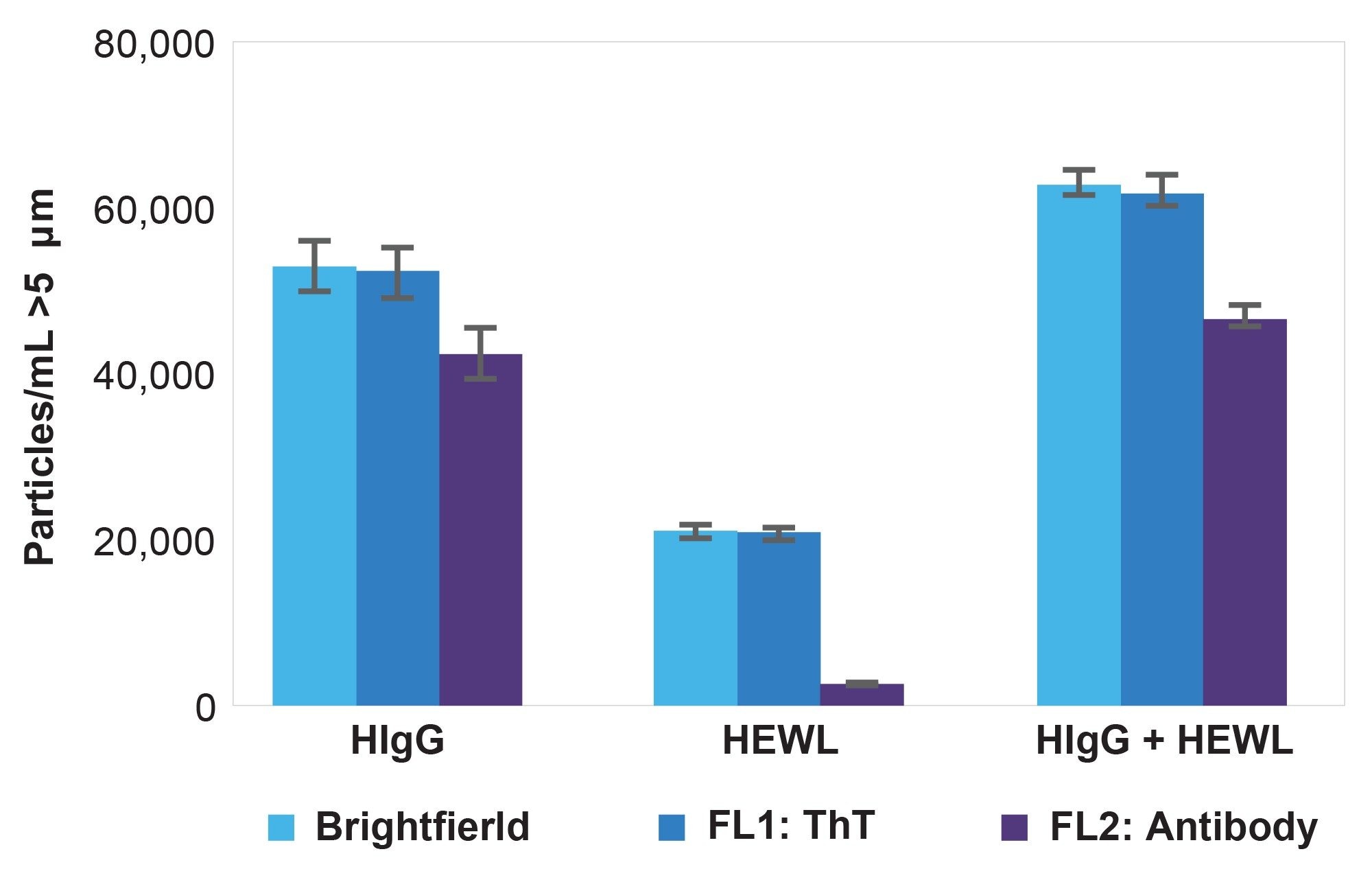
Quantitated total protein counts are measured in brightfield (all particles), FL1-ThT (all proteins), and FL2-Antibody (HIgG only) channels (Figure 4). Total fluorescent counts were calculated using appropriate thresholding in the expression engine function of Particle Vue Software. The data show that the Aura immunoassay correctly detects over 80% of HIgG protein aggregates > 5 µm present on the membrane, while delivering a 13% false positive rate, likely correlating with the overall specificity of the antibody to protein aggregates vs. native state proteins. Negative controls containing ETFE, antibody only, and PBS did not display significant values for FL2-Antibody signal despite the presence of particles in the brightfield (ETFE, and Antibody only) as well as FL1-ThT (antibody only) (data not shown). Thioflavin-T is a highly sensitive and specific protein dye which reliably stains all proteins aggregates (>99% staining efficiency for aggregates ≥5 µm). The difference in the number of FL1-ThT and FL2-Antibody positive particles in the HEWL group corresponds to the decrease in the respective groups in the HIgG + HEWL group. These data show that the Aura immunoassay is capable of accurate and precise protein labeling and these data confirm specific labeling of Human IgG protein aggregates with primary and secondary antibody, as well as their detection with the Aura System.
Conclusion
The Aura System is capable of specifically detecting different protein aggregates on a membrane using fluorescent antibody-based labeling. Additionally, this assay discerns between two different antibodies (HIgG and BIgG), which is useful for antibody co-formulation applications and assessing drug product vs carrier protein aggregates.
Powered with imaging in brightfield, as well as two fluorescent channels, the Aura System can help bridge the gap with common assays and subvisible particle stability. Custom fluorescence channels are optional to adapt to your fluorescent assay needs.
Featured Products
720009624, October 2026


