• 응용 자료

Removal of Polyethylene Glycol 400 (PEG 400) from Plasma Samples Using Mixed-Mode Solid-Phase Extraction

Removal of Polyethylene Glycol 400 (PEG 400) from Plasma Samples Using Mixed-Mode Solid-Phase Extraction

  • Jonathan P. Danaceau
  • Erin E. Chambers
  • Kenneth J. Fountain
  • Waters Corporation

Abstract

This application note describes a simple, quick, and elegant method for the removal of high concentrations of PEG from plasma samples through the use of Oasis mixed-mode cation-exchange (MCX) SPE, eliminating matrix effects often seen in early time points of pharmacokinetic studies.

Benefits

  • Removal of PEG 400 from plasma
  • Elimination of PEG 400 related ion suppression in early PK time points
  • Quick and easy sample preparation and cleanup without method manipulation

Introduction

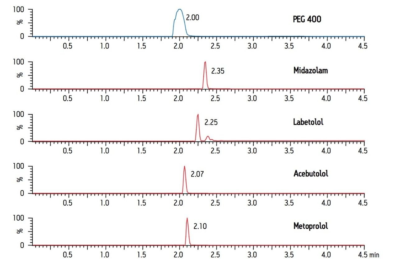
Pharmaceutical dosing vehicle excipients such as PEG 400 are often added to formulations to facilitate dissolution in dosing media. Unfortunately, these compounds can cause significant matrix effects, typically ion suppression, in LC-MS/MS analyses. The use of fast LC gradients, common in drug discovery, can result in co-elution of target analytes with these compounds. Early pharmacokinetic (PK) time points, when the concentration of these excipients is elevated, can be particularly troublesome as this co-elution has been shown to be a major contributing factor to excipient related ion suppression.1-3 Several approaches have been investigated to attempt to minimize this problem, including LC gradient manipulation, 2,3 alternative analytical column choices,1 different sample preparation strategies,1-3 sample dilution,5 and even the development of a novel formulating agent.4 While some of these strategies have been successful, they each have their limitations. Clearly, the choice of formulation excipient is often beyond the control of the analyst. LC optimization, either by gradient or mobile phase manipulation, or by alternative column selection can solve this problem for some analytes, but not always, and requires additional method development. In addition, extensive manipulation of LC conditions is not always conducive to a high-throughput screening environment. One group of researchers stated that “…if a more elegant solid-phase extraction (SPE) method…could be developed, an effective cleanup could possibly still be achieved.”3

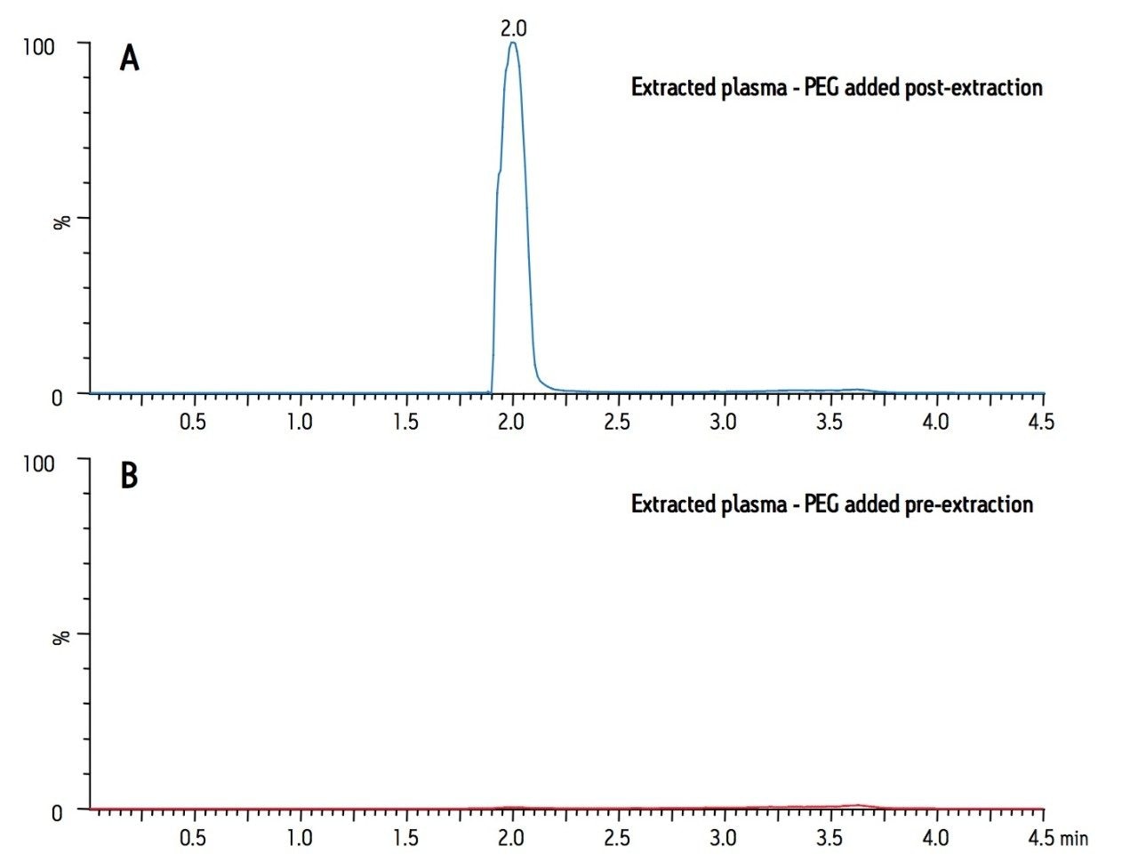
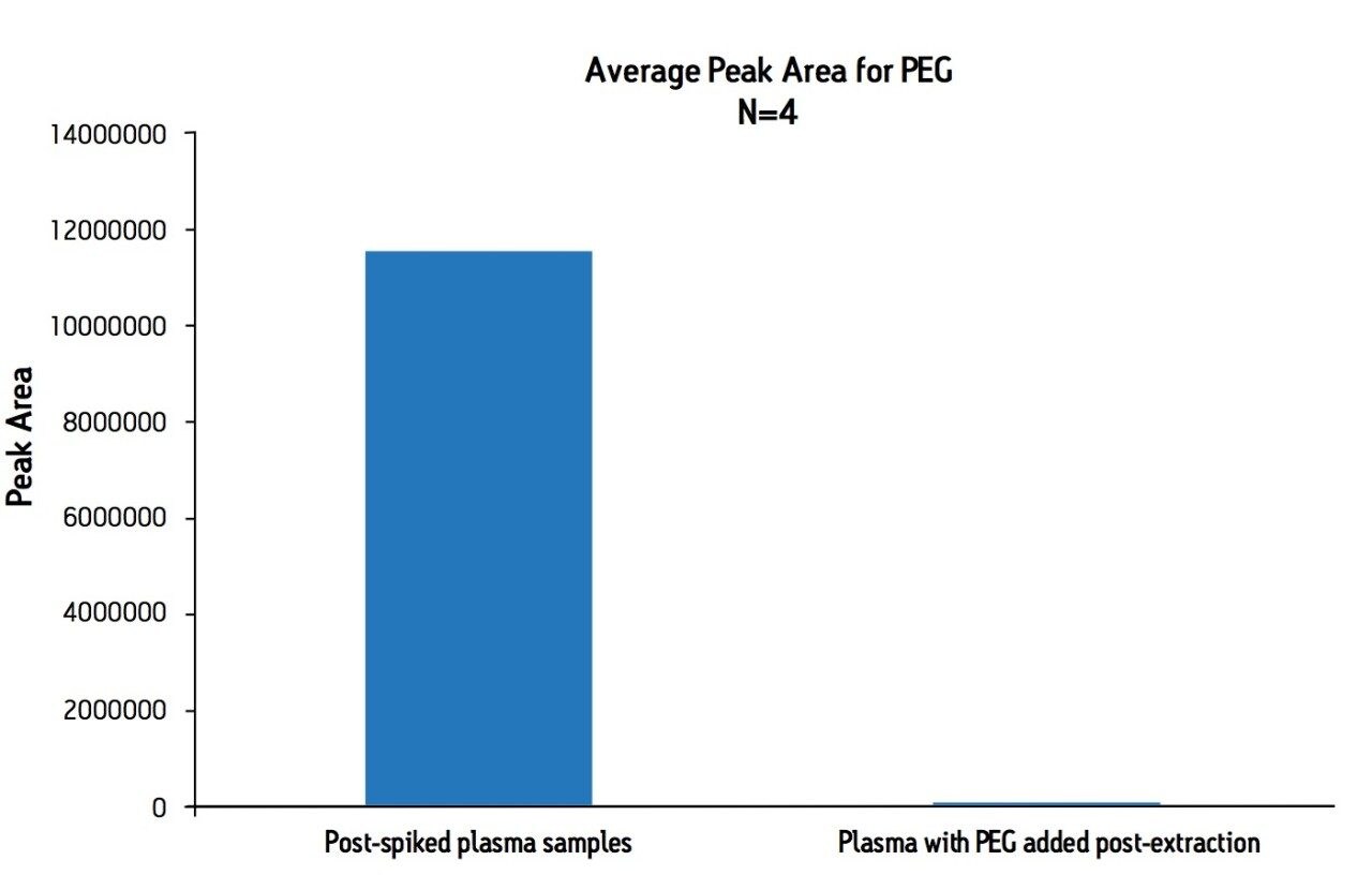
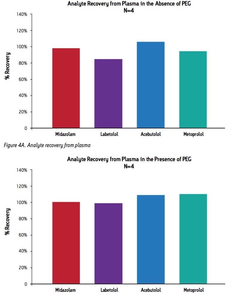
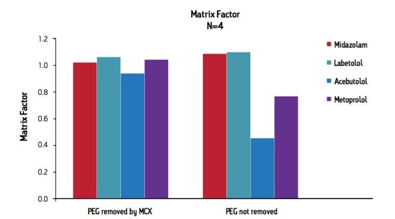
This work represents a fast, simple, and elegant solution to this problem through the use of Oasis mixed-mode cation-exchange (MCX) SPE. Basic analytes are bound to the sorbent by strong cation-exchange, and non-ionic excipients such as PEG, binds via a reverse phase mechanism, and can be selectively removed before analyte elution.

Experimental

Sample Preparation

A basic drug mixture was prepared that consisted of acebutolol, metoprolol, labetolol, and midazolam. This was either added to plasma samples prior to extraction to achieve plasma concentrations of 200 ng/mL, or used to post-spike blank extracted plasma eluates for recovery calculations. PEG 400 was either added to plasma prior to extraction at a concentration of 5 mg/mL or used to post-spike extracted blank plasma eluates for recovery determination.

Samples were extracted according to the basicprotocol for Oasis MCX. Briefly, 0.5 mL of rabbit plasma was acidified with 0.5 mL of 4% H3PO4. (Note: 0.5 mL of plasma was used in this study; smaller volumes may also be used). After conditioning MCX plates with 1.0 mL MeOH and 1.0 mL H2O, acidified samples were loaded onto  the sorbent bed. Loaded wells were then washed with 1.0 mL of 2% formic acid, followed by 2 x 1.0 mL of MeOH. The samples were then eluted with 2 x 250 μL of MeOH containing 5% NH4OH. “Post spike” (PS) samples were prepared by extracting blank rabbit plasma and post-spiking the final eluate with either the basic drug mix or a combination of the basic drug mix and PEG 400. “Extracted” samples were prepared by adding either 200 ng/mL basic drug mix or 200 ng/mL basic drug mix + 5 mg/mL PEG 400 to plasma prior to extraction.

Drug recovery and determination of PEG removal were determined by the following equation:

% Recovery = (peak area in extracted samples/peak area in post-spike samples) x 100%

For analysis of PEG removal, samples were diluted 1:200 in mobile phase to avoid contaminating the mass spectrometer with high concentrations of PEG 400.

SPE Conditions

SPE Plate:

Oasis MCX 96-well plate 30 μm (30 mg), Part number;186000248

LC Conditions

LC System:

Waters ACQUITY UPLC system

Detection:

Waters ACQUITY SQD

Vials:

2 mL 96-well collection plate; Part number WAT058958

Column:

ACQUITY UPLC BEH C18 1.7 μm, 2.1 x 50 mm, Part number:186002350

Column Temp.:

30 °C

Sample Temp.:

10 °C

Injection Volume:

10 μl

Flow Rate:

0.5 mL/min.

Mobile Phase A (MPA):

0.1% HCOOH

Mobile Phase B (MPB):

Acetonitrile containing 0.1% HCOOH

The initial mobile phase conditions were 95:5 MPA:MPB. Following a 0.5 min hold, MPB was increased to 90% over 2.5 min. The percentage of MPB was then returned to 5% and held for 2 minutes to re-equilibrate the column. The total run time was 5.0 min.

MS Conditions

MS System:

Waters ACQUITY SQD

Ionization Mode:

ESI Positive

Acquisition Range:

SIR

Capillary Voltage:

2.5 kV

Cone Voltage:

Compound specific (optimized for each analyte)

Desolvation Gas:

700 L/min.

Cone Gas:

0 L/min.

Data Management

Chromatography:

MassLynx V4.1 SCN714 and MS Software

Results and Discussion

Figure 1 shows the chromatography of PEG 400 and the 4 test compounds. The wide peak for PEG actually represents the combined TICs of the molecular ions from the most abundant components of PEG 400. This includes individual PEGs with masses of 370, 414, 458, 502, 546, 590, and 634. The test compounds (acebutolol, metoprolol, labetolol, and midazolam) are represented by SIR traces of the MH+ ion, with the exception of midazolam, in which the MH+ is combined with (MH-35)+. Note that acebutolol and metoprolol both co-elute with the PEG peak, which could make them especially vulnerable to ion suppression from residual PEG present in plasma samples. Figure 2 shows two chromatograms of PEG. Panel A indicates the level of PEG that would be present in an extracted plasma sample without removal by SPE. It was prepared by spiking PEG into the final plasma eluate post-extraction. Panel B is a chromatogram from a plasma sample containing 5 mg/mL PEG that was extracted using the standard MCX protocol, and demonstrates the nearly complete removal of PEG 400 from the plasma sample. Figure 3 shows the average PEG peak area from each sample group in the experiment and demonstrates that greater than 99% of the PEG 400 was removed by the MCX extraction procedure.

Figure 1. PEG 400 and basic drug mix chromatography.

Analyte recovery was calculated according to the formula described in the methods section. Figure 4A shows that all analytes had recoveries greater that 80% using the basic MCX protocol. The average analyte extraction recovery was 95% for the four analytes. Figure 4B shows that the presence of 5 mg/ml PEG 400 in the plasma did not affect analyte recovery. The average recovery in the presence of PEG was 103.6%.

Figure 4B. Analyte recovery in the presence of PEG

One of the key goals of PEG 400 removal from plasma is the elimination of excipient related ion suppression. Figure 5 is a summary of the matrix factor data calculated according to the equation below, as described in a recent AAPS article6.

Matrix factor = (peak response in presence of PEG/ peak response in absence of PEG)

The data in panel A are from plasma samples in which PEG had been removed by Oasis MCX extraction. The data in panel B are from plasma samples in which PEG had been added after sample extraction, and indicate the expected results if PEG had not been removed by sample preparation. This figure shows that if PEG is not removed, acebutolol and metoprolol suffer significant ion suppression as indicated by Matrix Factors of 0.45 and 0.77, respectively. These data clearly show that ion suppression is eliminated, even for those analytes that co-elute with the PEG peak, when PEG is removed using Oasis MCX SPE plates. Final matrix factors after PEG removal are 0.93 and 1.04, for acebutolol and metropolol, respectively.

Conclusion

Oasis MCX 96-well extraction plates represent a simple, quick, and elegant method for the removal of high concentrations of PEG from plasma samples, eliminating matrix effects (and, thus, ion suppression) often seen in early time points of pharmacokinetic studies. Furthermore, this problem is solved without any additional chromatographic method development, sample dilution, or other time consuming or cumbersome procedures, which makes it suitable and broadly applicable for a high-throughput environment.

References

  1. Tong X, Wang J, Zheng S, Pivnichny J, Griffin P, Shen X, Donnelly M, Vakerich K, Nunes C, and Fenyk-Melody J. Effect of signal interference from dosing excipients on pharmacokinetic screening of drug candidates by liquid chromatography/mass spectrometry. Anal Chem 2002 74:6305-6313.
  2. Weaver R and Riley R. Identification and reduction of ion suppression effects on pharmacokinetic parameters by polyethylene glycol 400. Rapid Comm Mass Spec 2006 20:2559-2564.
  3. Shou W and Naidong W. Post-column infusion stude of the ‘dosing vehicle effect’ in the liquid chromatography/tandem mass spectrometric analysis of discovery pharmacokinetic samples. Rapid Comm. Mass Spec 2003 17:589-597.
  4. Temesi D, Law B, and Howe N. Synthesis and evaluation of PEG414, a novel formulating agent that avoids analytical problems associated with polydispersive vehicles such as PEG400. J. Pharmaceutical Sciences 2003 92(12):2512-2518.
  5. Larger P, Breda M, Fraier D, Hughes H, and James C. Ion-suppression effects in liquid chromatography-tandem mass spectrometry due to a formulation agent, a case study in drug discovery bioanalysis. J. Pharm. Biomed. Anal. 2005 39:206-216.
  6. Bansal S and DeStefano A. Key elements of bioanalytical method development validation for small molecules. The AAPS Journal 2007 9(1):E109-E114.

720004127, October 2011

맨 위로 돌아가기 맨 위로 돌아가기