• アプリケーションノート

Charge Reduction Electron Transfer Dissociation (crETD) and Ion Mobility for Deconvolution of Protein Mass Spectra and Conformational Analysis

Charge Reduction Electron Transfer Dissociation (crETD) and Ion Mobility for Deconvolution of Protein Mass Spectra and Conformational Analysis

  • Frederik Lermyte
  • Jonathan P. Williams
  • Jeffery M. Brown
  • Esther M. Martin
  • Frank Sobott
  • Waters Corporation

Abstract

This application note describes a method of extensive charge reduction of native protein complexes on unmodified SYNAPT G2 and SYNAPT G2-S Ion Mobility Mass Spectrometers.

Benefits

  • Extensive charge reduction via ion/ion chemistry leads to increased peak separation and simplified spectral assignment of native protein complexes on unmodified instruments
  • Concomitant ETD fragmentation in exposed terminal regions provides sequence information
  • Ion mobility allows structural characterization of charge-reduced species

Introduction

The production of high-mass, multiply charged ions via electrospray ionization (ESI) permits mass spectrometric analysis of native protein complexes.1,2 However, given the relatively broad peaks typical in native MS, mass and charge state assignment can be challenging, particularly for heterogeneous samples.3 Furthermore, a correlation exists between protein charge state and conformation, and lower charge states are often believed to resemble the native (solution) state more closely.4 Therefore, methods of reducing the gas-phase charge state offer analytical benefits. Previous efforts have generally either relied on ejection of highly charged monomers,3 causing significant disruption of the native structure, or have required instrument modification.5

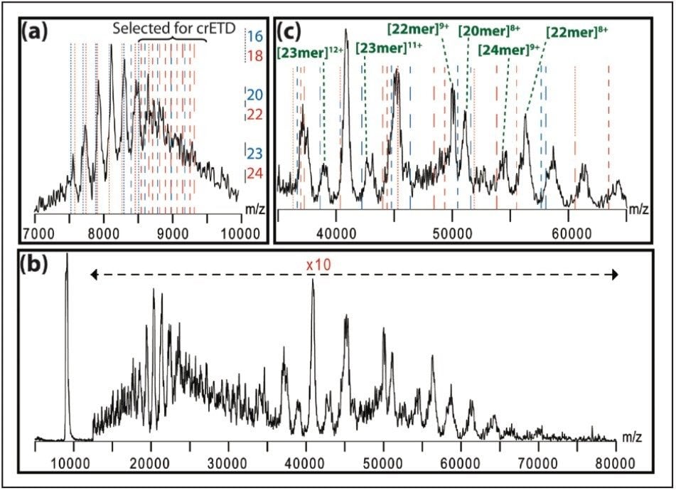
Here, we describe a method of extensive charge reduction of native protein complexes on unmodified SYNAPT G2 and SYNAPT G2-S Ion Mobility Mass Spectrometers. This results in increased peak separation and simplified spectral assignment, as illustrated in Figure 1, but also allows us to investigate electrostatic effects on protein folding state by using ion mobility. Concomitantly, some ETD fragments, originating from surface-exposed terminal regions, are also observed, providing sequence information.

Figure 1. (a) Native ESI spectrum of αB-crystallin (red/blue dashed lines: calculated m/z values); (b) crETD spectrum, with increased peak spacing; (c) magnification of (b), with some species annotated.

Experimental

Sample description

Proteins were dissolved at a concentration of 1 mg/mL in 100 mM aqueous ammonium acetate (pH = 6.8) and desalted twice using Bio-Rad Micro Bio-Spin 6 columns.

Method conditions

Samples were introduced via direct infusion. Both 1,4-dicyanobenzene and p-nitrotoluene were used as ETD reagents, yielding similar results.

MS conditions

MS systems:

SYNAPT G2 and SYNAPT G2-S

Capillary voltage:

1.0 kV

Trap pressure:

8e-2 mbar

Trap DC bias:

11 V (45 V in Mobility mode)

Transfer pressure:

6e-3 mbar (2e-2 mbar in Mobility mode), transfer CE 5 V

Results and Discussion

Besides the polydisperse αB-crystallin, shown in Figure 1, we also tested this method on five monodisperse complexes: hemoglobin (64 kDa), concanavalin a (103 kDa), alcohol dehydrogenase (ADH, 148 kDa), pyruvate kinase (233 kDa), and L-glutamic dehydrogenase (GDH, 336 kDa). As shown in Figure 2, this leads to the observation of extensive charge reduction in all cases.

For all proteins, it was necessary to increase the post-ETD acceleration voltages in order to observe the low charge states. Both the trap DC bias and the transfer collision energy proved effective for this (in TOF mode), as shown in Figure 3. ETD fragmentation in the N-terminal region of ADH is visible in the low (600–3200) m/z region in this figure, as reported earlier.6,7

Figure 3. ADH spectra (26+ precursor isolated in quadrupole) showing the effect of varying degrees of post-ETD ion acceleration, with ETD fragments in red.

Finally, the IM capabilities of the SYNAPT G2 and SYNAPT G2-S instruments can be used to study the conformation of the charge-reduced complexes, and investigate the relation between the charge and folding state. In order to minimize disturbance of the protein structure, the trap DC bias and transfer CE were kept at 45 V and 2 V, respectively. However, even under these conditions, charge reduction of 26+ ADH down to the 15+ charge state was observed, as shown in Figure 4. The table in Figure 4b shows the calculated collision cross-section (CCS) of each observed charge state. Within the observed range, no major structural changes were observed, other than a gradual decrease in CCS due to reduced Coulombic repulsion. This is consistent with earlier work carried out on a modified ion mobility mass spectrometer.5

Figure 4. (a) crETD of 26+ ADH in Mobility mode, showing charge reduction down to the 15+ ion; (b) calculated collision cross-sections for the charge states observed in (a).

Conclusion

Extensive charge reduction of noncovalent protein complexes weighing up to several hundred kDa was demonstrated on unmodified SYNAPT G2 and SYNAPT G2-S instruments using standard ETD reagents. This method was used to partially deconvolute the very complex signal generated by a large, polydisperse assembly, allowing identification of the major species present. Concomitant top-down fragmentation provides sequence information. In combination with ion mobility, the effect of charge state on the global conformation of proteins and protein complexes can be studied.

References

  1. Fenn JB, Mann M, Meng CK, Wong SF, Whitehouse CM. Electrospray ionization for mass spectrometry of large biomolecules. Science. 1989 Oct 6;246(4926):64–71. 
  2. Benesch JLP, Ruotolo BT, Simmons DA, Robinson CV. Protein complexes in the gas phase: technology for structural genomics and proteomics. Chem Rev. 2007 Aug;107(8):3544–67. 
  3. Aquilina JA, Benesch JL, Bateman OA, Slingsby C, Robinson CV. Polydispersity of a mammalian chaperone: mass spectrometry reveals the population of oligomers in alphaB-crystallin. Proc Natl Acad Sci U S A. 2003 Sep 16;100(19):10611–6. 
  4. Hall Z, Robinson CV. Do charge state signatures guarantee protein conformations? J Am Soc Mass Spectrom. 2012 Jul;23(7):1161–8. 
  5. Campuzano I, Schnier P. Coupling electrospray corona discharge, charge reduction and ion mobility mass spectrometry: From peptides to large macromolecular protein complexes. Int J Ion Mobil Spec. 2013;16(1):51–60. 
  6. Lermyte F, Konijnenberg A, Williams JP, Brown JM, Valkenborg D, Sobott F. ETD Allows for Native Surface Mapping of a 150 kDa Noncovalent Complex on a Commercial Q-TWIMS-TOF Instrument. J Am Soc Mass Spectrom. 2014 Mar;25(3):343–50. 
  7. Lermyte F, Sobott F. Electron transfer dissociation provides higher-order structural information of native and partially unfolded protein complexes. Proteomics. 2015 Aug;15(16):2813–22.

Solutions in practice

  • Lermyte F, Williams JP, Brown JM, Martin EM, Sobott F. Extensive Charge Reduction and Dissociation of Intact Protein Complexes Following Electron Transfer on a Quadrupole-Ion Mobility-Time-of-Flight MS. J Am Soc Mass Spectrom. 2015 Jul;26(7):1068–76.
  • Williams JP, Lermyte F, Anderson M, Jackson L, Giles K, Brown JM, Sobott F, Langridge J. Advances in Native Mass Spectrometry-Based Methods for the Analysis of Noncovalent Protein Complexes. Proceedings of the 62nd ASMS Conference on Mass Spectrometry and Allied Topics, Baltimore, Maryland. 2014 (P/N PSTR134803339).

Featured Products

720005484, November 2015

トップに戻る トップに戻る