
Investigating the Strength of the Sense and Antisense Oligonucleotide Interaction to Form siRNA Duplexes Using the BioAccord™ LC-MS System
Catalin Doneanu, Ying Qing Yu
Waters Corporation, United States
Published on August 03, 2026

Abstract
A hydrophilic interaction liquid chromatography (HILIC)-UV/MS assay was developed for the analysis of three distinct intact small interfering RNA (siRNA) duplexes under native mass spectrometry conditions. The mass spectrometric data revealed that weakly associated duplexes underwent more extensive in-source dissociation than their more tightly associated counterparts. These results demonstrate that the LC–UV/MS systems can provide valuable insights into the relative interaction strength between the sense and antisense strands that comprise siRNA duplexes.
Benefits
- A single HILIC-UV/MS assay was developed on the BioAccord electrospray ionization-time-of-flight (ESI-TOF) Mass Spectrometer to measure the neutral monoisotopic mass of several intact siRNA duplex molecules while preserving their higher-order structure and distinguishing weakly versus tightly associated duplexes
- HILIC provides strong retention for very polar siRNA species, enabling separation of duplexes from the corresponding single oligonucleotide strands and related impurities
- This HILIC LC-UV/MS assay avoids expensive and toxic reagents (such as hexafluoroisopropanol (HFIP) amines/modifiers) and supports both qualitative and quantitative characterization of siRNA therapeutics for identity, purity, and stability studies
Introduction
siRNA duplexes are double-stranded RNA molecules typically consisting of two 21–23 oligonucleotides named the sense (passenger) strand and the antisense (guide) strand. In therapeutic applications of the siRNA interference mechanism, siRNA duplexes are designed to target disease-associated genes at the mRNA level, reducing the production of harmful proteins without altering the underlying DNA sequence. This approach forms the basis of a few drugs recently approved by regulatory agencies, as highlighted by several oligonucleotide therapeutics reviews.1–4
The LC-UV/MS analysis of siRNA duplexes was described in two recent application notes from Waters Corporation5,6, and the general interest in these types of molecules is reflected by the introduction in 2024 of a siRNA duplex LC-MS standard oligonucleotide by Waters (p/n: 186010598).
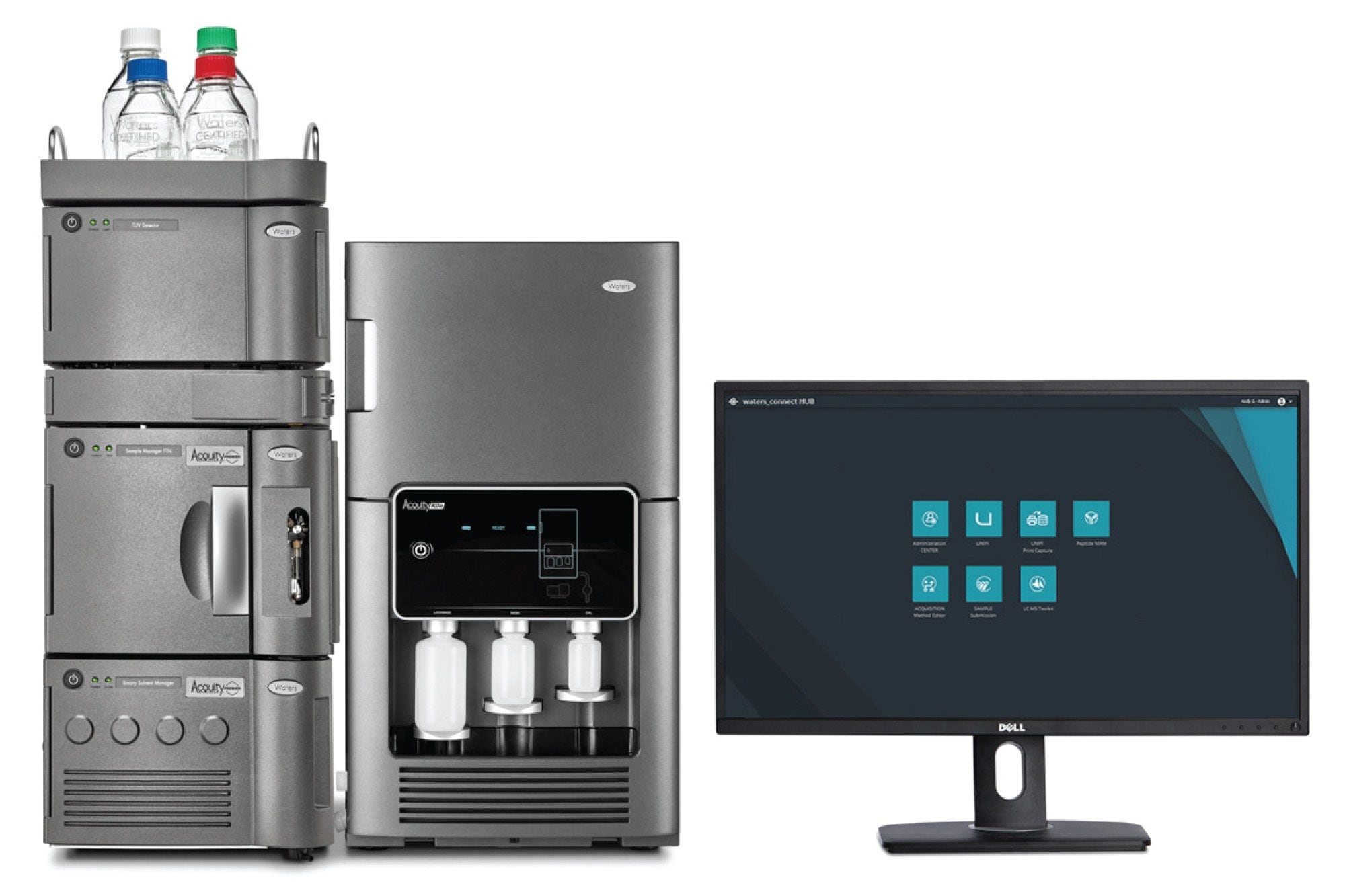
While ion-pairing reversed-phase (IP-RP) chromatography typically denatures non-covalent siRNA duplexes, it has recently been demonstrated that HILIC preserves the oligonucleotide structure7,8 , enabling native mass spectrometry experiments to be performed even in the presence of high organic solvent content. Here, the HILIC LC-UV/MS analysis of three different siRNA duplexes are described and the utility of the BioAccord LC-UV/MS TOF System are demonstrated for investigating the binding affinity of the sense and antisense nucleotide strands. Chromatographic separations were performed on the recently introduced ACQUITY™ Premier BEH™ Amide HILIC Columns9 , which feature a very inert internal column surface, specifically designed to minimize electrostatic interactions between very polar analytes and the metal surfaces located inside the UPLC™ columns.
Experimental
Reagents and Sample Preparation
LC-MS grade LiChropur™ ammonium acetate (catalog number 5.33004.0050) was purchased from MilliporeSigma (St Louis, MO) and acetonitrile (LC-MS grade, catalog number 34967-6XL) was obtained from Honeywell (Charlotte, NC). HPLC grade Type I deionized (DI) water was purified using a Milli-Q® system (Millipore, Bedford, MA).
Three siRNA duplexes containing a pair of sense and antisense oligonucleotides designed to target three different proteins were ordered from Abbexa (Sugar Land, TX): 1) catalogue number abx909935- a SEM1 duplex specific for the SEM1 subunit of the 26S proteasome mouse protein; 2) abx907084 – an axin interactor dorsalization-associated (AIDA) duplex targeting a human protein localized in the outer cellular membrane; 3) abx909453 – an ATPase PAAT duplex designed to bind a human mitochondrial enzyme (PAAT) that hydrolyzes ATP to regulate the function of ABC transporter proteins. Lyophilized duplexes (15 nanomoles each) were dissolved in DI water to prepare stock solutions with a concentration of 50 µM. The stock solutions were diluted ten fold with solvent A (75% acetonitrile, 10 mM ammonium acetate, pH 9.0) to prepare 5 uM siRNA duplex solutions which were analyzed via LC-UV/MS using 1 µL sample injections.
All HILIC-UV/MS datasets were acquired with waters_connect™ Software on the BioAccord LC-MS System.
|
LC-MS system: |
BioAccord LC-MS System with ACQUITY Premier UPLC System |
|
Column: |
ACQUITY Premier BEH Amide Column 1.7 µm, 130 Å, 2.1 x 50 mm, (p/n: 186009504) |
|
Column temperature: |
35 °C |
|
Flow rate: |
300 µL/min |
|
Mobile phases: |
Solvent A: 75% acetonitrile, 10 mM ammonium acetate, pH 6.8 Solvent B: 25% acetonitrile in DI water, 10 mM ammonium acetate, pH 6.8 |
|
Wash solvents: |
Purge solvent: 75% acetonitrile, 10 mM ammonium acetate, pH 6.8 Sample manager wash: 75% acetonitrile, 10 mM ammonium acetate, pH 6.8 Seal wash: 20% acetonitrile in DI water |
|
Detector: |
ACQUITY UPLC TUV Detector, 260 nm, 20 Hz data acquisition rate |
|
Sample temperature: |
6 °C |
|
Sample vials: |
LCMS Certified Maximum Recovery Vials (p/n: 186005663CV) |
|
Injection volume: |
1 µL |
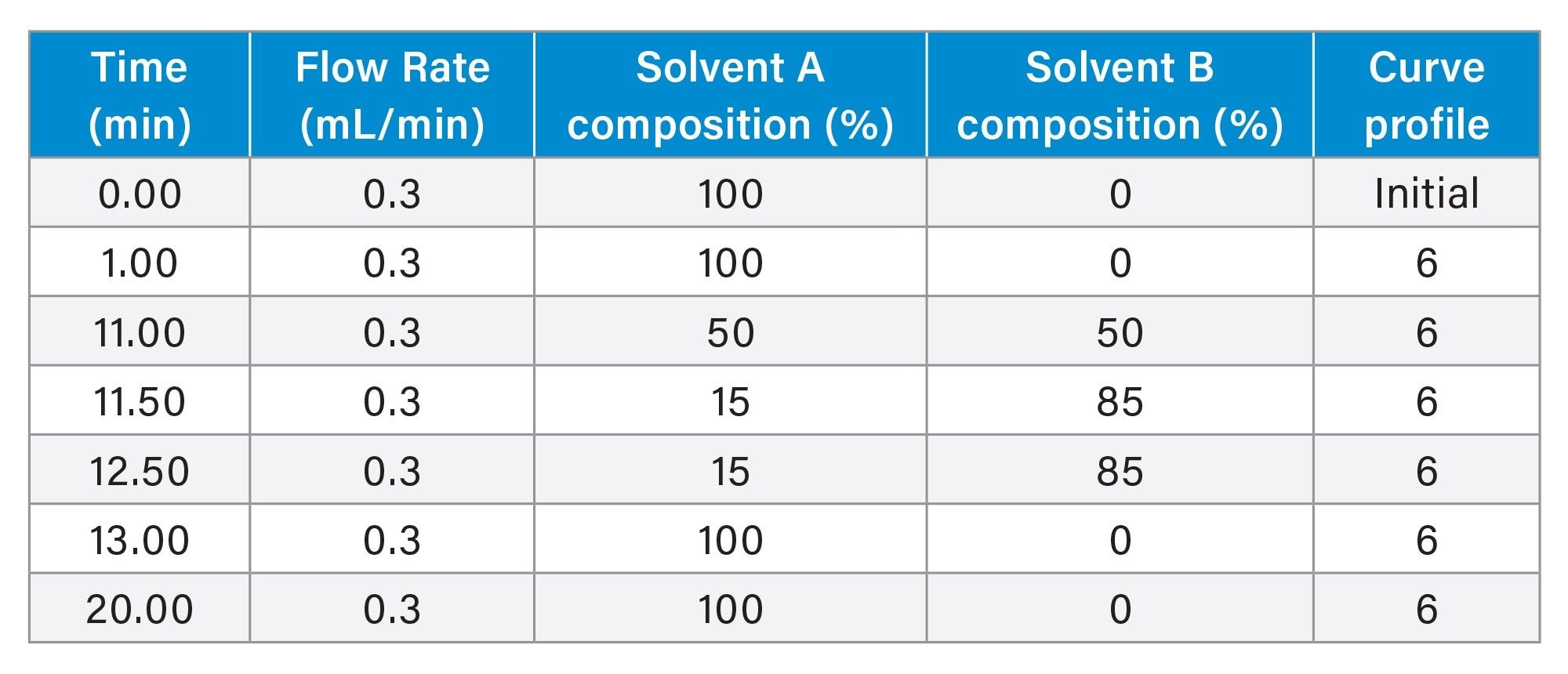
Gradient Table
|
Acquisition mode: |
Full scan MS mode |
|
Ionization mode: |
ESI(-) |
|
Capillary voltage: |
0.8 kV |
|
Cone voltage: |
20 V |
|
Source temperature: |
120 oC |
|
Desolvation temperature: |
500 oC |
|
Desolvation gas (N2) pressure: |
6.5 bar |
|
TOF mass range: |
400–5000 |
|
Acquisition rate: |
2 Hz |
|
LockMass: |
waters_connect LockMass Solution (p/n: 186009298) |
|
Data acquisition software: |
waters_connect Software |
|
Data processing software: |
waters_connect Software |
Results and Discussion
Native mass spectrometric analysis of intact siRNA duplexes following HILIC separation has recently been reported.5–8 Notably, the high concentrations of organic solvents commonly employed in HILIC mobile phases, including acetonitrile, methanol, and even 1,1,1,3,3,3-hexafluoroisopropanol (HFIP), do not appear to disrupt the native duplex structure of oligonucleotides. This contrasts with proteins and peptides, which generally undergo significant denaturation under similar solvent conditions. Nucleic acid duplexes have remarkable stability under conditions that would typically induce denaturation of protein/peptide-based biomolecules, because they are stabilized by different forces than proteins/peptides. The key reason is that RNA duplex stability is dominated by Watson–Crick base pairing, base stacking interactions, and electrostatic screening by cations, whereas protein/peptide native structures depend heavily on the hydrophobic interactions. These hydrophobic interactions are strongly weakened in organic-rich solvents, which is why proteins often denature in HILIC-like mobile phases. In addition, the siRNA duplex melting temperature (Tm), rather than solvent composition alone, determines whether the sense and antisense oligonucleotide strands remain annealed. In HILIC separations, intact siRNA duplexes can remain stable provided the chromatographic temperature stays below the duplex melting temperature. Previous HILIC studies have shown intact duplex peaks under suitable conditions and strand dissociation only when column temperature is approaching Tm.5–7
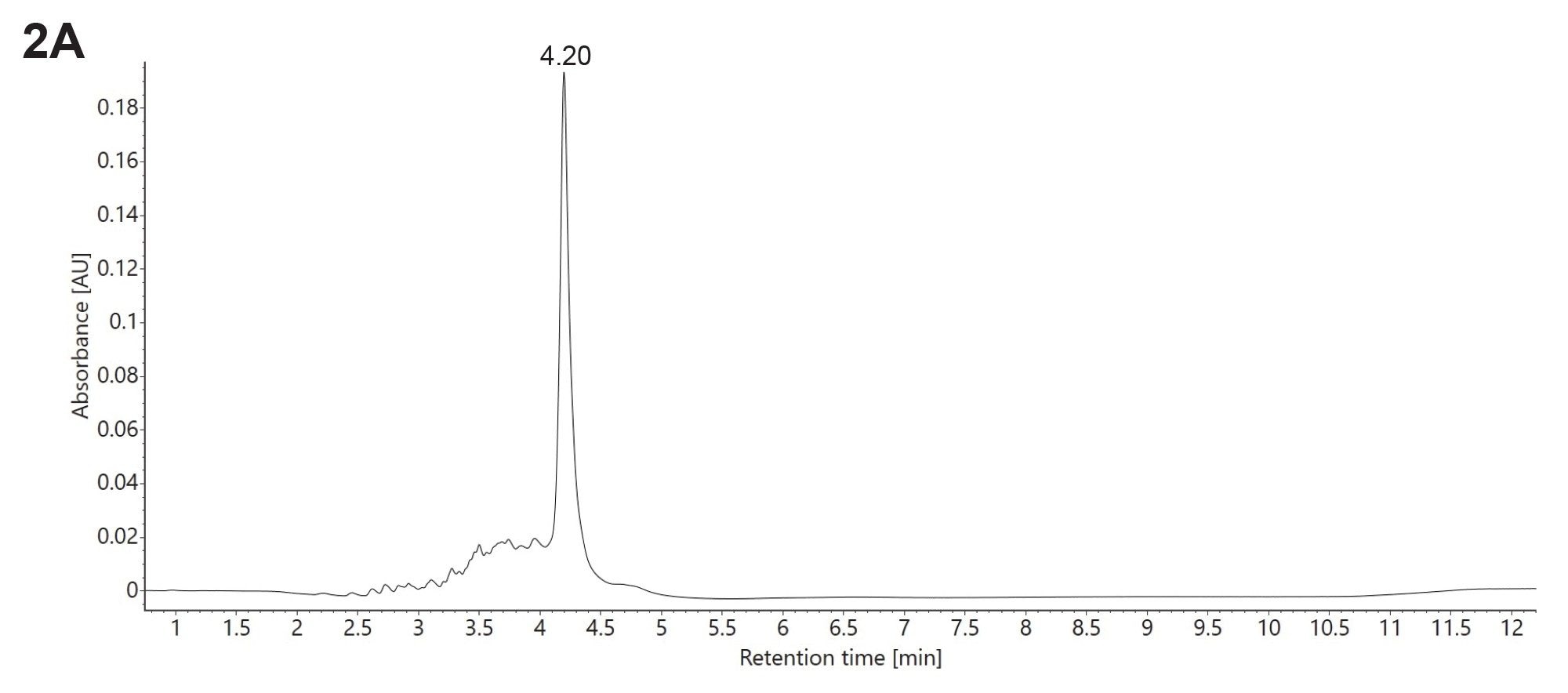
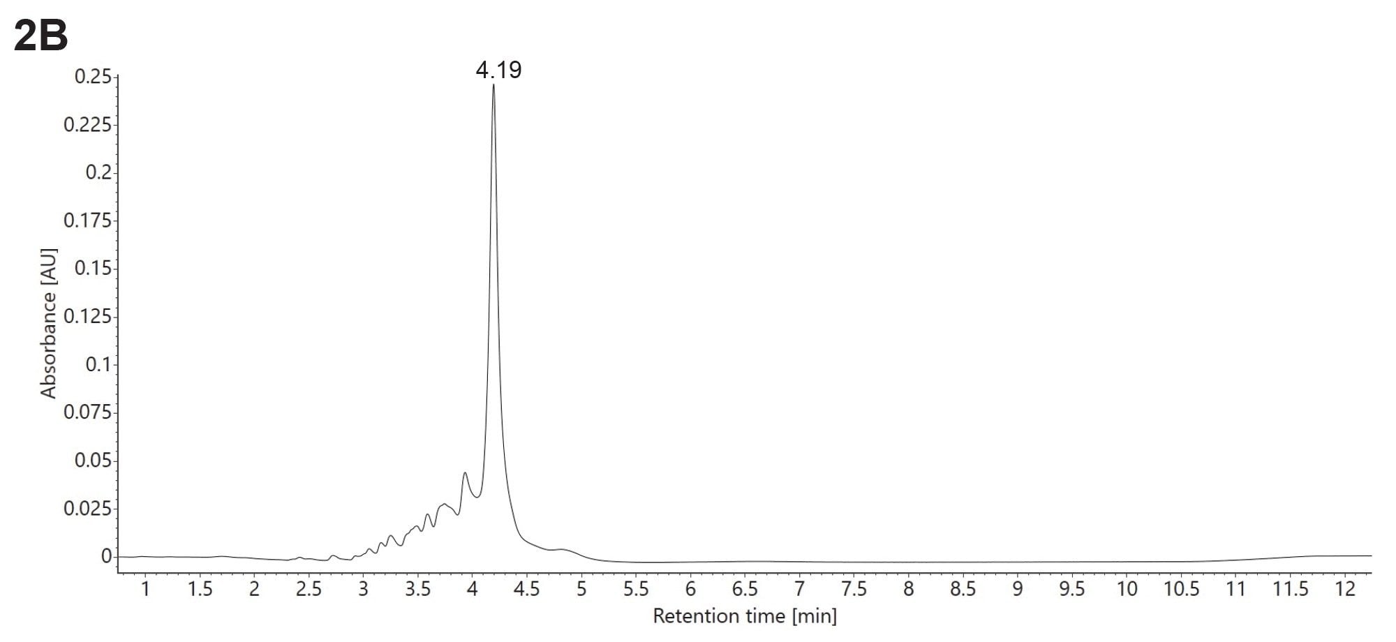
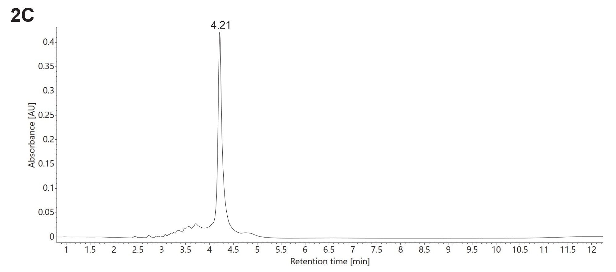
All three siRNA duplexes investigated in this study produced a single dominant chromatographic peak when analyzed by HILIC at a column temperature of 35 °C, well below their respective Tms, as illustrated by the UV chromatograms shown in Figure 2A–C. The samples were analyzed using identical chromatographic conditions, including a gradient reaching 50% eluent B after 10 minutes. Despite differences in the sequence and composition of the sense and antisense strands, the intact siRNA duplexes exhibited very similar retention times. As evident from the individual UV chromatograms, each sample contained numerous oligonucleotide-related impurities, reflecting the limited purification of the constituent sense and antisense strands prior to duplex formation. Nevertheless, the intact siRNA duplex remained the predominant species detected in all three samples. Collectively, these findings demonstrate the robustness of the HILIC method in preserving the native structure of siRNA duplexes across a range of oligonucleotide sequences and compositions.
The combined ESI-MS spectra obtained for the intact siRNA duplexes following HILIC-MS analysis are shown in Figure 3A–C. In all three cases, the native siRNA duplex was detected within a narrow charge-state distribution, spanning only three charge states, from [M−5H]⁵⁻ to [M−7H]⁷⁻. This limited charge-state range is in agreement with the presence of a compact, native-like, duplex structure in the gas phase. In contrast, IR-RP chromatography performed at the same column temperature (35 °C) resulted in complete denaturation of each siRNA duplex. Under these conditions, only the individual sense and antisense strands were detected as separate chromatographic peaks, while no intact duplex signal was observed (data not shown).
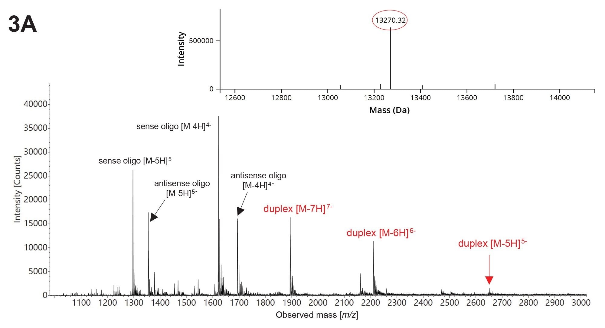
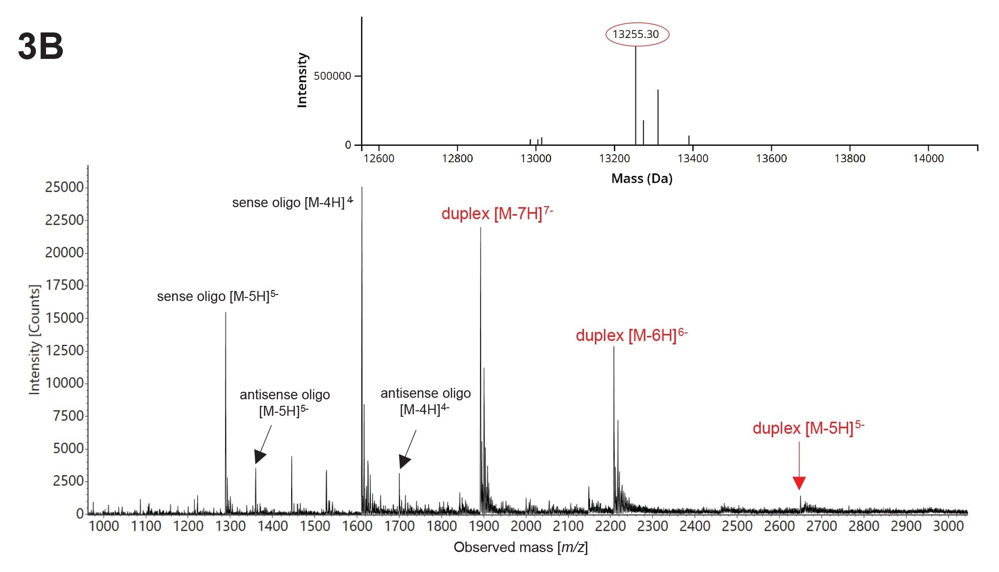
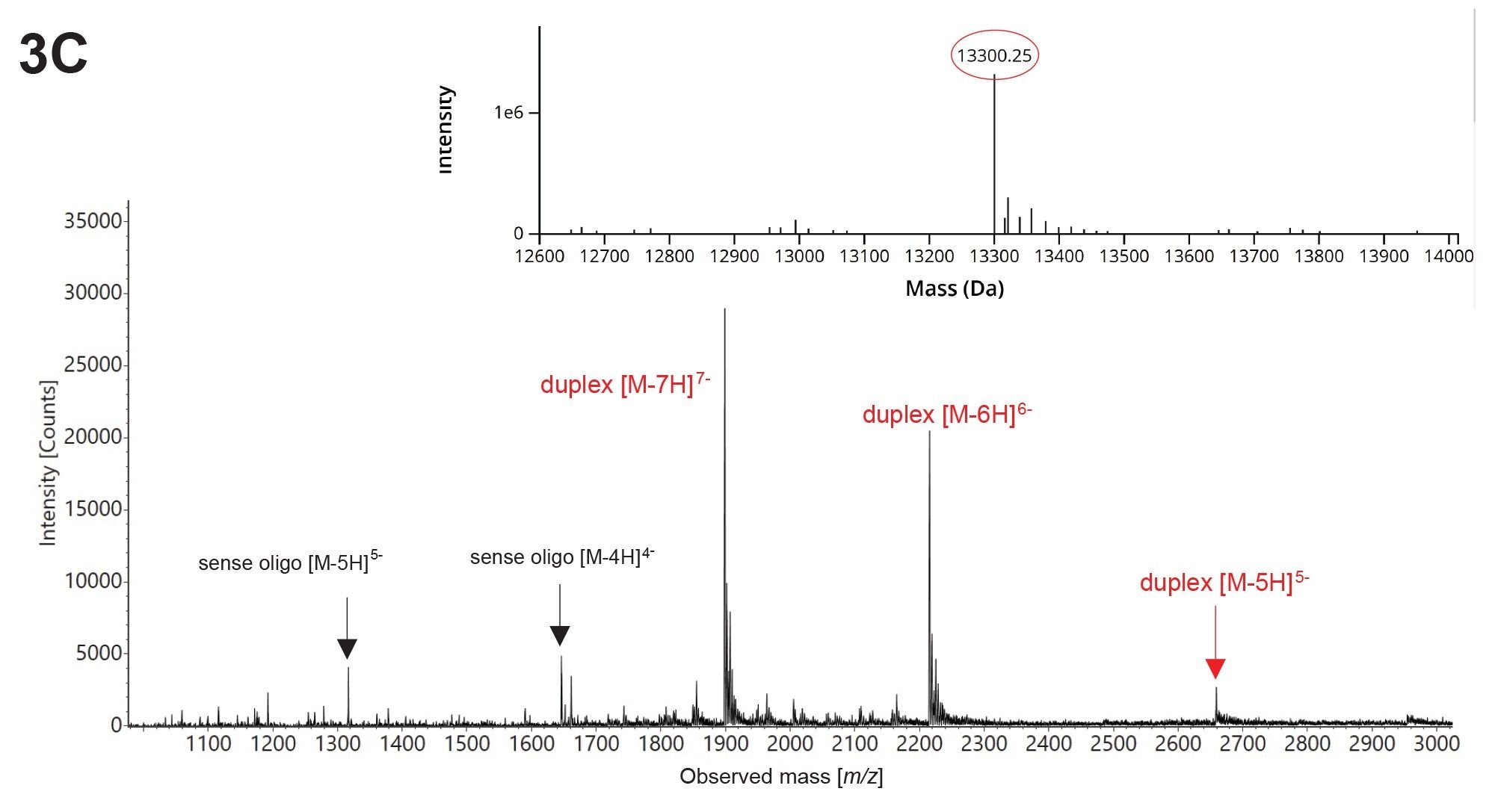
Because siRNA duplexes can undergo partial dissociation during the electrospray ionization process, several key source parameters on the BioAccord System were optimized to enhance the gas-phase stability of non-covalent RNA complexes. These parameters included the capillary voltage, cone voltage, desolvation temperature, and desolvation gas flow. The optimized conditions, summarized in the experimental section, were employed for the acquisition of the spectra presented in Figure 3A–C. Deconvolution of the siRNA duplex ESI-MS spectra was performed in the INTACT Mass App v1.910 using the BayesSpray deconvolution algorithm11 , enabling determination of the neutral monoisotopic mass of each duplex. The corresponding deconvolved spectra are displayed as insets in Figures 3A–C.
Two siRNA duplexes, the SEM1 duplex (Figure 3A) and the AIDA duplex (Figure 3B), underwent partial dissociation during the electrospray ionization process, despite the use of optimized BioAccord ESI source parameters. Consequently, signals corresponding to the individual sense and antisense oligonucleotide strands were also observed in the mass spectra as shown in Figures 3A and 3B. Assignment of the sense and antisense charge states were based on the average molecular weights of the corresponding oligonucleotides, as provided by the siRNA duplex manufacturer (Abbexa, Sugar Land, TX). However, no sequence information or nucleotide composition data were supplied for the individual siRNA duplex components. These results indicate that the interactions between the sense and antisense strands in the SEM1 and AIDA duplexes were not sufficiently strong to maintain complete duplex integrity during the transition from solution to the gas phase. In contrast, the ATPase PAAT duplex, shown in Figure 3C, exhibited substantially greater gas-phase stability, as evidenced by the high abundance of intact duplex ions corresponding to the native charge states (-5 to -7) observed in the ESI-MS spectrum. The in-source dissociation of this siRNA duplex is minor, as indicated by the weak signals of the quadruply charged states of its sense and antisense strands shown in Figure 3C. Because identical ESI source parameters were used on the BioAccord ESI-TOF Mass Spectrometer for the analysis of all three duplexes, the enhanced gas-phase stability of the ATPase PAAT duplex is likely attributable to stronger interactions between its sense and antisense strands relative to those of the SEM1 and AIDA duplexes. These findings demonstrate that the BioAccord LC-UV/MS Platform can be used to assess the relative strength of strand association within siRNA duplexes by monitoring their stability during electrospray ionization and transfer to the gas phase.
The results presented here clearly suggest that the HILIC LC-UV/MS instrument can provide valuable insights into the relative interaction strengths of siRNA duplex components.
Conclusion
HILIC can maintain siRNA duplexes and other higher-order oligonucleotide structures during chromatographic separation, enabling direct analysis of intact duplexes rather than inferring duplex behavior from single strands. A HILIC LC-UV/MS setup simultaneously acquires UV and MS signals for quantitative and qualitative characterization, using MS-friendly mobile phases that avoid toxic modifiers and ion-pairing reagents and support flexible switching between different LC methods and mobile phases. When deployed within the Waters ecosystem, these capabilities are integrated into a unified, compliant data and workflow environment that streamlines method development, review and transfer across labs.
References
- Goyon, A.; Yehl, P.; Zhang, K. Characterization of Therapeutic Oligonucleotides by Liquid Chromatography, J Pharm Biomed Anal, 2020, 182, 113105, doi: 10.1016/j.jpba.2020.113105.
- Vinjamuri, B.P.; Pan, J.; Kawakami, J.; Peng, P. A Review on Commercial Oligonucleotide Drug Products, J Pharm Sci, 2024, 113, 1749–1768, doi/10.1021/acs.analchem.5c06656.
- Togawa, H.; Yamaguchi, T.; Kawakami, J.; Obica, S. Current Trends in Liquid Chromatography-Mass Spectrometry Analysis of small Interfering rNAs: A short review, Anal Chem, 2026, 98, 10895–10912, doi/10.1016/j.chroma.2025.466550.
- Bardawil, S.A.; Barthelemy, P.; Ferey, L. Advances in the Analysis of Therapeutic Oligonucleotides with Chromatography Coupled to Mass Spectrometry, Anal Chem, 2026, 98, 10895–10912, doi/10.1021/acs.analchem.5c06656.
- Hirschberger, R.; Rühl, M.; Fox, J.; Yu, Y.Q.; Berger, S. Characterization and Sequencing of Duplex siRNA Using the BioAccord LC-MS System and VION IMS QTof Mass Spectrometer in Combination with the waters_connect CONFIRM Sequence App. (2024), Waters Application Note. 720008654.
- Fekete, S.; Araya, M.; Addepalli, B.; Lauber, M.A. Studying Mobile Phases for HILIC UV/MS Analysis of Oligonucleotide Therapeutics including an siRNA Duplex, a Lipid Conjugated ASO, and a CRISPR sgRNA. (2024), Waters Application Note. 720008602.
- Huang, M.; Xu, X.; Qiu, H.; Li, N. Analytical Characterization of DNA and RNA oligonucleotides by Hydrophilic Interaction Liquid Chromatography-Tandem Mass spectrometry, J Chrom A, 2021, 1648, 462184, doi 10.1016/j.chroma.2021.462184
- Doneanu, C.E.; Yu, Y.Q.; Fredette, J.; Warren, W.J.; Chen, W. HILIC as an Alternative Separation Mode for Intact Mass Confirmation of Oligonucleotides on the BioAccord System. (2021), Waters Application Note. 720007395.
- Berthelette, K.D.; Kalwood, J.; Haynes, K. Development and Optimization of a HILIC MS Separation of 17 Free Amino Acids Using an XBridge Premier BEH Amide Column. (2023), Waters Application Note. 720008010.
- Shion, H.; Boyce, P.; Berger, S.J.; Yu, Y.Q. INTACT Mass – A Versatile waters_connect™ Application for Rapid Mass Confirmation and Purity Assessment of Biopharmaceuticals. (2022), Waters Application Note. 720007547.
- Skilling, J.; et al. Beyond MaxEnt Deconvolution: Increasing the Fidelity and Universal Applicability of Mass Spectral Deconvolution Routines for Biomolecules with the Application of Bayesian Probability Theory. (2020), Waters Poster. 720003756.
Featured Products
720009523, July 2026


