Fast, flexible platforms for peptide quantification are needed, particularly for a discovery setting. This type of methodology would be especially advantageous in the case of amyloid beta (aβ) peptides. The deposition/formation of insoluble aggregates, or plaques, of aβ peptides in the brain is considered to be a critical event in the progression of Alzheimer’s Disease (AD) and thus has the attention of many researchers.
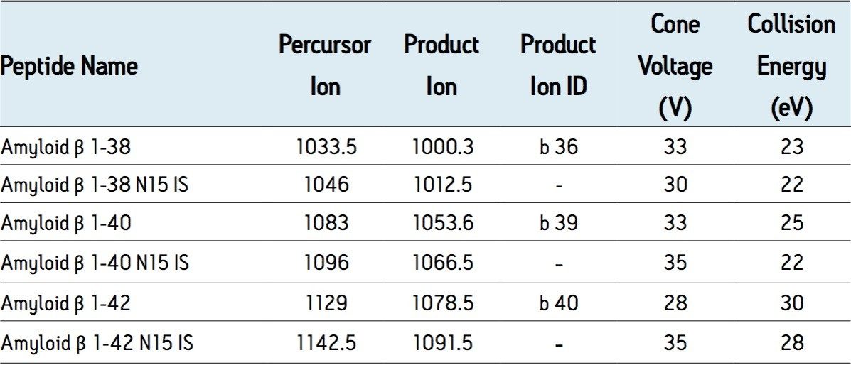
Historically, quantification of aβ peptides in biological fluids has relied mainly on the use of immunoassays, such as ELISA. These assays are time consuming and expensive to develop, labor intensive, are subject to cross reactivity and an individual assay is required for each peptide. In order to meet the throughput requirements and constant flow of demands for new peptide methods in a discovery setting, there is a need for a highly specific yet flexible methodology based on an LC-MS/MS platform. In this work, this platform is coupled with selective sample preparation for the simultaneous quantitation of multiple aβ peptides. This work focuses on methods for the 1-38, 1-40, and 1-42 aβ peptides, in support of preclinical studies.
Development of a bioanalytical method for these peptides is further complicated by their propensity for aggregation, formation of oligomers, poor solubility, nonspecific binding, and hydrophobicity.
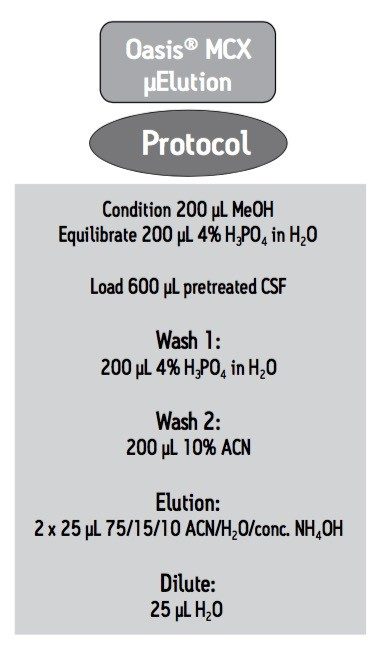
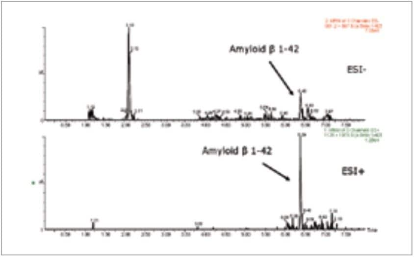
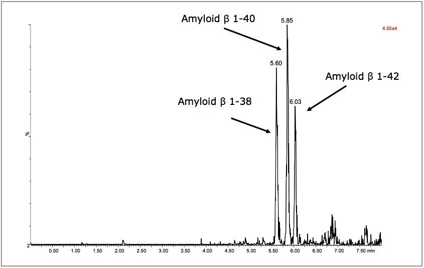
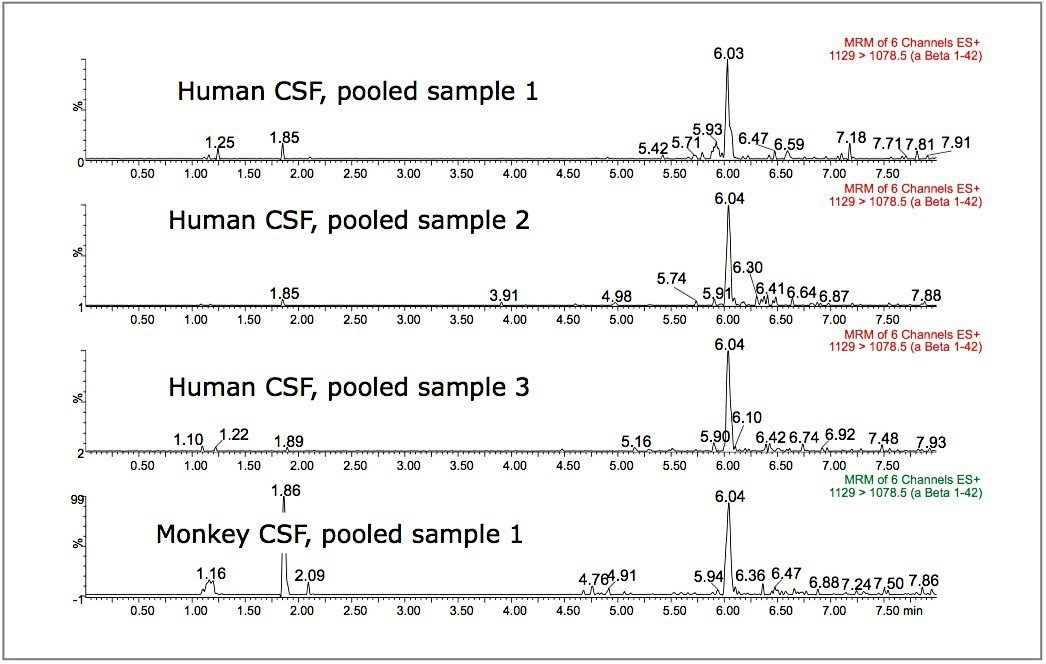
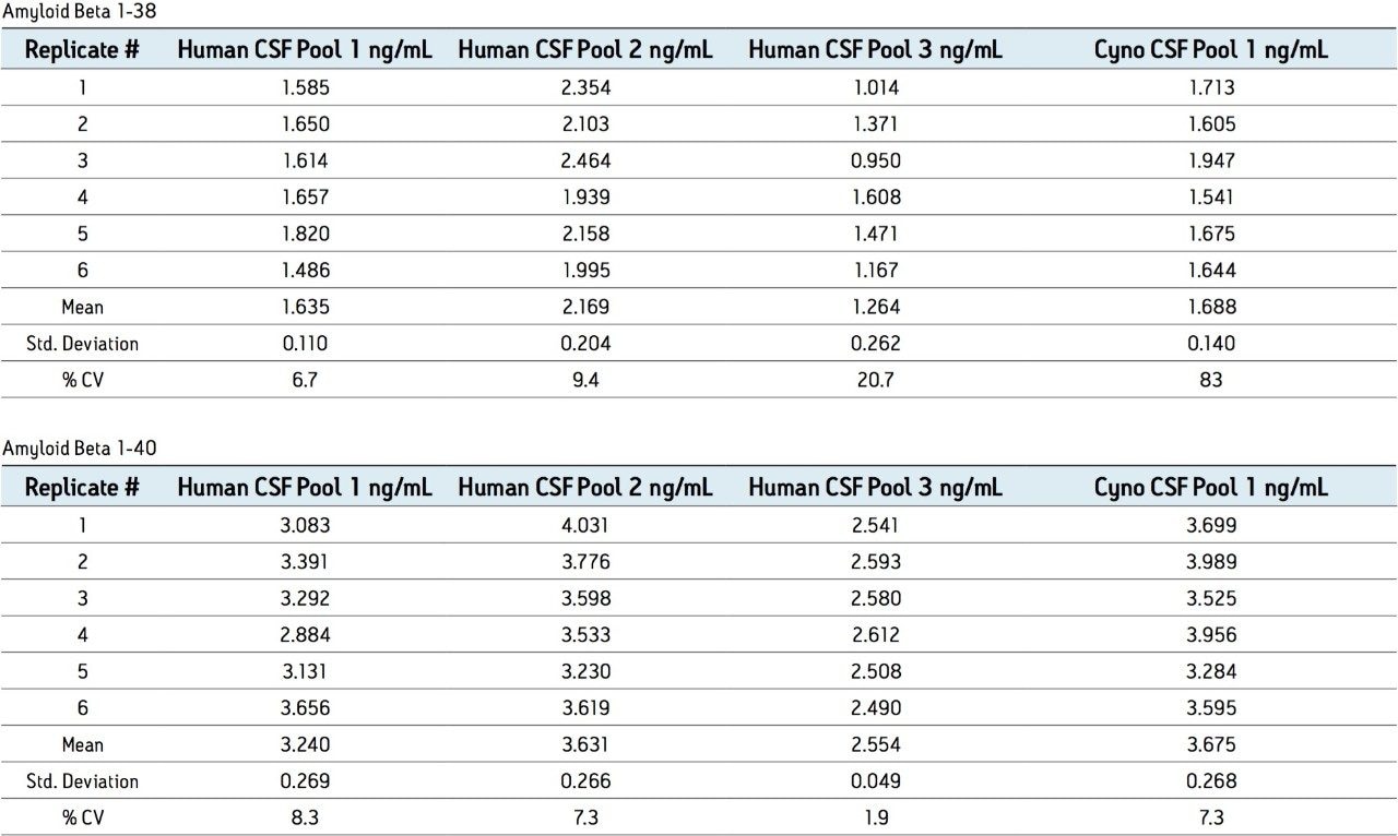
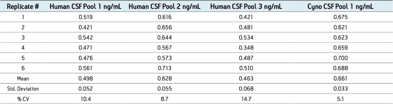
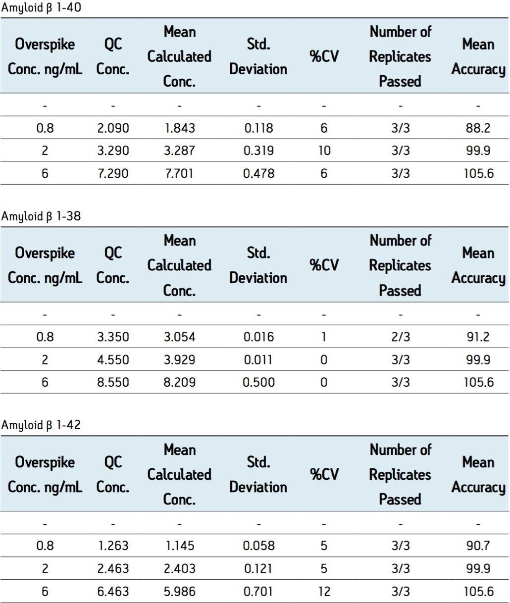
As aβ peptides may be present at very low concentrations, we developed a solid-phase extraction (SPE) sample preparation protocol to enrich the amyloid beta fraction in CSF. The SPE method concentrates the sample to improve detection limits while eliminating matrix interferences and optimizing solubility of the aβ peptides in the mass spectrometer injection solution. A high throughput, high resolution UPLC-MS/MS quantitation method was also developed.
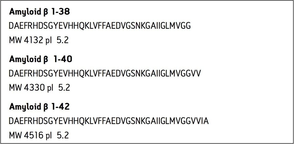
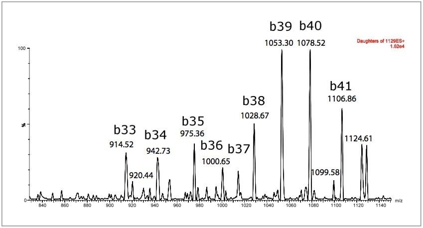
This work focuses on the development of UPLC, MS, and selective SPE sample preparation methods for the 1-38, 1-40, and 1-42 fragments of APP, in support of preclinical studies. Sequence, pI and molecular weight (MW) information for these peptides is shown in Figure 1. The use of a single, high throughput assay for multiple aβ peptides- without time consuming immunoprecipitation steps was developed and validated. The speed, selectivity, and specificity of this technique for simultaneously quantitating multiple aβ peptides in CSF are demonstrated.