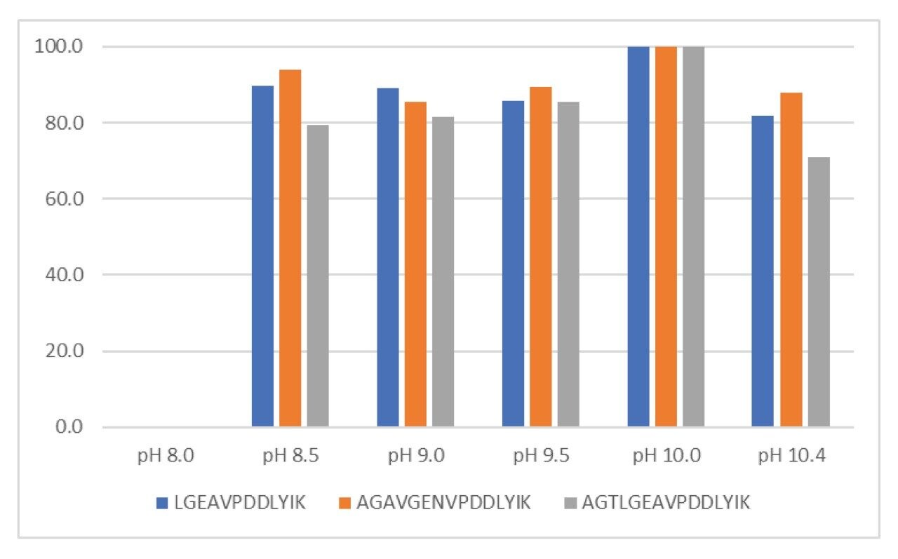
Quantification and identity testing are an important part of vaccine substance and product batch release. Traditional methods can be non-selective, time consuming to develop, and may have poor multiplexing capabilities. GARDASIL 9 is a nonavalent vaccine composed of nine different HPV L1 protein antigens that provide broad protection against HPV subtypes responsible for increased cervical cancer and genital wart risk. In this work, a complete sample preparation workflow (including adjuvant desorption, tryptic digestion, and LC-MS/MS quantitative monitoring) has been leveraged to measure GARDASIL 9 components via the surrogate peptide method. We were able to simultaneously quantify all nine HPV strain specific L1 proteins with high reproducibility in one LC-MS/MS method over the concentration range of 10–200 μg/mL.
GARDASIL 9 is a nonavalent vaccine composed of HPV L1 protein antigens which form virus like particles (VLPs). These antigens protect against nine HPV strains which account for 80–90% of cervical cancers and 90% of genital warts.1,2 An important step in the vaccine production process is quality control of vaccine substance and product batches. Batch release testing includes sterility, potency, purity, antigen quantification, and identity to name a few. Traditional methods for quantification and identity testing include total protein assays and ELISA. These assays have limitations which include non-specificity (or unknown specificity or cross reactivity), are time consuming to develop, and have poor multiplexing capabilities. There is therefore an opportunity to leverage mass spectrometry as a tool for both antigen quantification and identity testing. Mass spectrometry allows for the simultaneous and selective determination and quantification of type specific L1 proteins. In this work, we have developed a complete sample preparation workflow encompassing desorption of HPV L1 proteins from aluminum adjuvant, and tryptic digestion of proteins for the quantification of GARDASIL 9 HPV L1 antigens via the surrogate peptide method. This workflow, including mass spectrometric analysis, enabled the highly reproducible quantification of these peptides over the range of 10–200 μg/mL which easily covers the expected HPV L1 protein concentration range of 40–120 μg/mL in the GARDASIL 9 vaccine product.
Each HPV L1 protein was diluted to 750 µg/mL (30 µg) and denatured with RapiGest SF surfactant (Waters, P/N 186001861), followed by reduction with dithiothreitol and alkylation with iodoacetamide. Samples were digested with trypsin (Promega, P/N V5111) using a 1:10 trypsin:protein ratio (w/w) and quenched with formic acid. Peptide mapping using a LC-MSE method was performed by injecting 10 µL of sample onto an ACQUITY UPLC I-Class PLUS System coupled with a Xevo G2-XS QTof Mass Spectrometer.
Two microliters of blanks, standards, quality control, or vaccine product samples were added to a 16 µL aqueous solution of 100 µg/mL bovine serum albumin (Sigma, P/N 05470), respectively. 2 µL of internal standard was added to all samples except blanks. 20 µL of 0.1% (w/v) RapiGest SF surfactant (Waters, P/N 186001861) prepared in 100 mM Tris HCl, pH 10.0 was added to each sample. Samples were incubated for 10 minutes at 80 °C, then centrifuged for 5 minutes at 2000 x g to pellet adjuvant. 32 µL of sample was carefully removed from the pelleted adjuvant and neutralized to pH 8.0 with 1.2 µL of 10% hydrochloric acid (v/v). Samples were digested with 0.3 µg of trypsin (Promega, P/N V5111), (1:10 trypsin:protein ratio) at 37 °C overnight. The digestion reaction was quenched and RapiGest was precipitated with 2 µL of 20% formic acid in water (v/v) for 15 minutes at 37 °C. Samples were centrifuged at 1800 x g for 15 minutes at 10 °C to pellet RapiGest. 30 µL of each sample was transferred to a QuanRecovery 96-well plate and 5 µL was injected onto an ACQUITY UPLC I-Class PLUS System coupled to a Xevo TQ-S micro Mass Spectrometer for LC-MS/MS analysis.
LC system: |
ACQUITY UPLC I-Class PLUS (Fixed Loop) |
MS system: |
Xevo G2-XS QTof Mass Spectrometer, ESI+ |
Sample plates: |
QuanRecovery with MaxPeak High Performance Surfaces Vials |
Column(s): |
ACQUITY UPLC PREMIER Peptide BEH C18, 300 Å, 1.7 µm, 2.1 x 100 mm |
Column temp.: |
60 oC |
Sample temp.: |
10 oC |
Injection volume: |
10 µL |
Flow rate: |
0.2 mL/min |
Mobile phase A: |
0.1% Formic acid in water |
Mobile phase B: |
0.1% Formic acid in acetonitrile |
Gradient: |
1–35% B in 50 minutes at 0.2 mL/min followed by column wash and equilibration |
Ionization mode: |
ESI+ |
Capillary voltage: |
1.2 kV |
Cone voltage: |
40 V |
Source temp.: |
150 oC |
Desolvation temp.: |
350 oC |
Cone gas flow: |
20 L/Hr |
Desolvation gas flow: |
600 L/Hr |
Informatics system for data collection and processing: |
UNIFI (v.1.9.4) |
LC system: |
ACQUITY UPLC I-Class PLUS (Fixed Loop) |
MS system: |
Waters Xevo TQ-S micro Mass Spectrometer, ESI+ |
Sample plates: |
QuanRecovery with MaxPeak High Performance Surfaces 96-well plates |
Column(s): |
ACQUITY UPLC PREMIER Peptide BEH C18, 300 Å, 1.7 µm, 2.1 x 100 mm |
Column temp.: |
60 oC |
Sample temp.: |
10 oC |
Injection volume: |
5 µL |
Flow rate: |
0.4 mL/min |
Mobile phase A: |
0.1% Formic acid in water |
Mobile phase B: |
0.1% Formic acid in acetonitrile |
Gradient: |
10–15% B in 1 minute, 15–30% B in 3 minutes followed by column wash and equilibration. 7-minute run time. |
Ionization mode: |
ESI+ |
Capillary voltage: |
1.2 kV |
Cone voltage: |
20 V |
Source temp.: |
150 oC |
Desolvation temp.: |
600 oC |
Cone gas flow: |
100 L/Hr |
Desolvation gas flow: |
1000 L/Hr |
MS data collection: |
MassLynx v4.2 |
Informatics for data processing: |
TargetLynx XS v4.2 |
Characterization of HPV L1 proteins via a peptide mapping approach helps to select peptide candidates for quantitation and may also identify the presence of protein backbone modifications such as deamidation and oxidation. The complete workflow for protein analysis and quantification has been described previously for Fc containing proteins in application note 720006969EN and is used here with minor modifications.3 Peptide mapping experiments were performed for all nine HPV L1 proteins present in GARDASIL 9. As an example, shown in Figure 1, excellent fragmentation data confirms the HPV 18 peptide FSLDLDQYPLGR. This peptide was chosen as a surrogate of HPV 18 on the basis of intensity, low likelihood of modifications, and quantitative reproducibility. MRM transitions for each surrogate peptide were identified and optimized via the MassLynx-Skyline interface workflow.4
Briefly, after surrogate peptides are identified via peptide mapping experiments, retention times of these peptides are identified by monitoring precursor masses of each peptide using the optimized chromatographic method. The most intense charge state for each peptide precursor is chosen and all b and y ion product ions for the selected peptide are comprehensively examined to identify the most intense MRM transitions. The top MRM transitions are further optimized for collision energy and a final MS method is generated with these optimal parameters. MRM transitions used for quantification can be viewed in Table 1.
Column selection is an important part of the LC method development process. The ACQUITY PREMIER Peptide BEH C18 300 Å Column utilizing HPS (High Performance Surfaces) Technology was found to be optimal for the quantification of HPV L1 proteins. In Figure 2, a comparison of a standard column and a PREMIER Column is shown.5 Both columns were passivated with 10 injections of a high concentration sample for passivation, followed by 5 analyte injections. The PREMIER Column with MaxPeak HPS Technology enabled faster column passivation and provided a 2x increase in peak area over the standard column. This peak area increase allows lower limits of quantification and enhances reproducibility of peptide measurements.
Efficient adjuvant desorption is key to the success of a quantitative measurement of antigen concentration in adjuvanted vaccine products. Proteins can bind to aluminum adjuvants via electrostatic forces, hydrophobic interactions, hydrogen exchange via Van der Waals forces, and ligand exchange.6 Ligand exchange provides the tightest binding of proteins and results from interactions between surface hydroxyl groups of adjuvant and protein antigen phosphate groups. GARDASIL 9 is adjuvanted to the proprietary amorphous aluminum hydroxyphosphate sulfate (AAHS) which has surface hydroxyl and phosphate groups.7 This vaccine is formulated at approximately pH 7.0 and AAHS has a point of zero charge at this pH.8 The antigen HPV L1 protein VLPs have isoelectric points ranging from 8.1 to 8.6 and are therefore positively charged in the formulated vaccine product.9,10
We have experimentally determined that surfactants and high pH are needed in order to disrupt this adsorption, completely desorb proteins from the AAHS adjuvant, as well as begin to dissociate the VLPs themselves.11 We have used the MS-friendly surfactant RapiGest to denature the HPV L1 proteins which was required to enable complete desorption. Our adjuvant desorption protocol also utilizes high pH to fully switch the charge of the HPV L1 proteins from positive to negative. Shown in Figure 3, pH 10.0 achieved the highest level of protein desorption from the adjuvant. Important to note, very little protein desorption was achieved at pH 8.0 where the proteins are expected to be positively charged. This result indicates that electrostatic repulsion was required to adequately disrupt binding and lead to protein desorption.
Complete digestion of proteins is essential for accurate, sensitive, and reproducible quantification via the surrogate peptide method. In order to ensure complete digestion, digestion parameters such as enzyme:substrate ratio and digestion time must be examined and optimized. To 
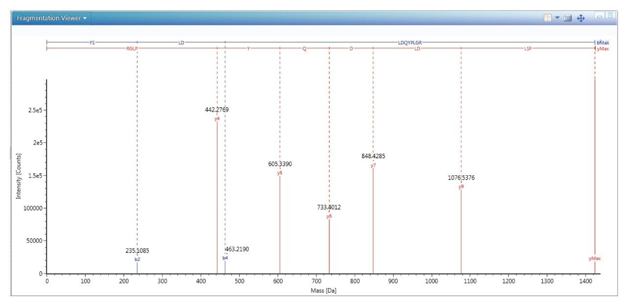
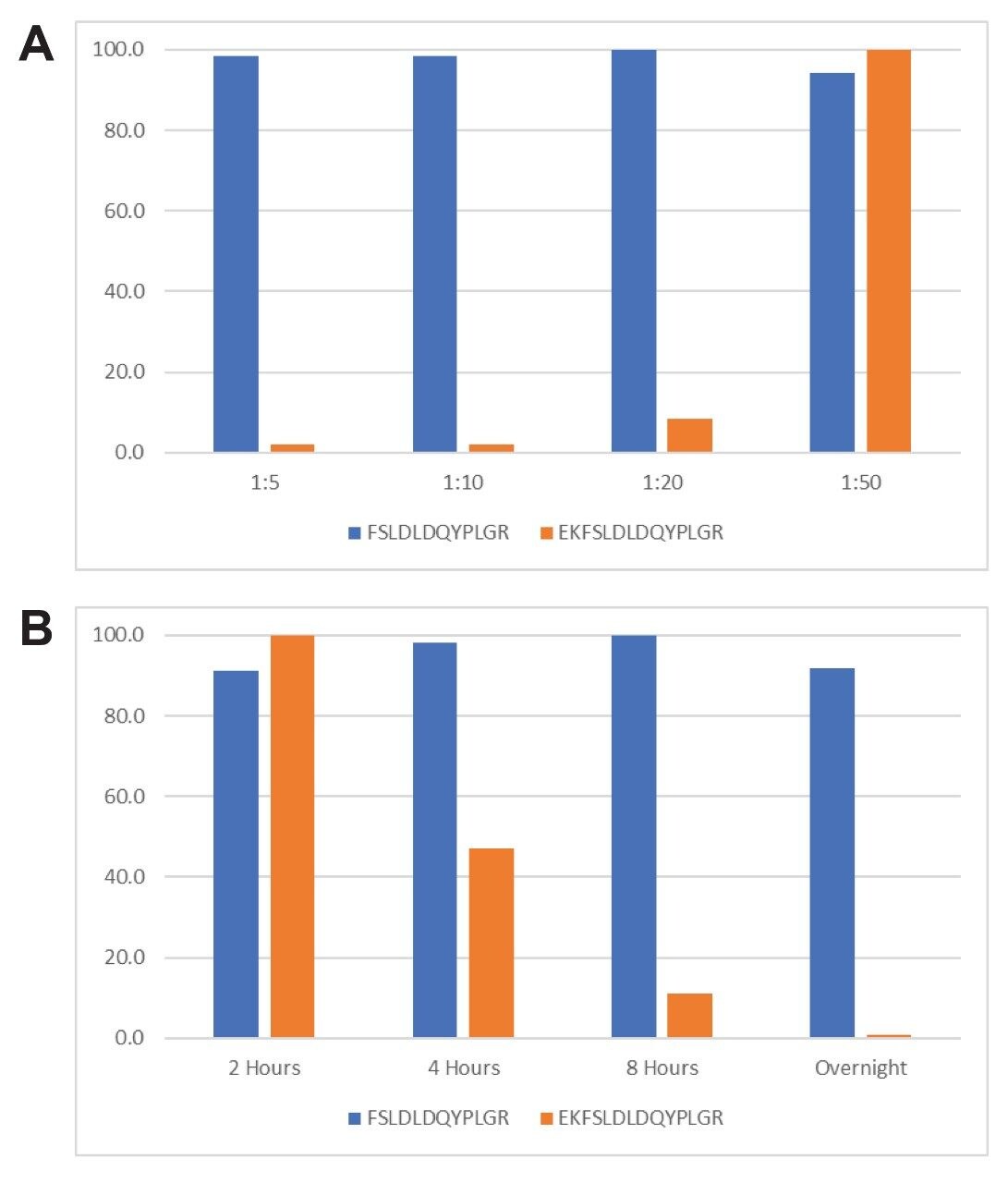
measure digestion completeness, the peptide FSLDLDQYPLGR and the peptide EKFSLDLDQYPLGR with one missed cleavage were monitored and quantified.
The ratio of trypsin to protein was optimized based on the overnight digestion results at various enzyme:substrate ratios. The results of these experiments are shown in Figure 4, panel A. At the trypsin:protein ratio of 1:10 and 1:5, the maximum intensity of peptide FSLDLDQYPLGR and the minimum intensity peptide EKFSLDLDQYPLGR were obtained, suggesting a more complete digestion of the HPV L1 proteins was achieved. A 1:10 ratio was chosen to use in the rest of the experiments to reduce the autolysis of the trypsin due to the need of high concentrations. Shown in figure 4, panel B, digestion time was evaluated. A digestion time of 8 hours to overnight resulted in the most complete digestion of the HPV L1 proteins. Overnight digestion was chosen due to completeness of digestion and workday convenience.
Standard curves, QCs, and vaccine product were prepared over three days to validate inter- and intra-day assay precision and accuracy. Calibration curves were linear for HPV 16 over the range of 20–200 µg/mL. The remaining HPV proteins were linear over the range of 10–100 µg/mL (Table 2). Low, medium, and high QCs were prepared, and the CVs for the accuracy and precision of QCs were <8% (Table 3). Shown in Table 4, measurement of all nine HPV L1 proteins was highly reproducible over three preparations and three days of measurements with CVs <8%. In most cases, LC-MS/MS measurements in this study agreed with the labelled values in general, although some values were higher. These results indicate that this assay is fit for purpose for the quantification of HPV L1 proteins in GARDASIL 9.
A mass spectrometry assay for the simultaneous quantification of HPV L1 protein antigens in GARDASIL 9 was developed. Combined with optimized sample preparation, proteins were quantified by LC-MS/MS with high accuracy and precision.
720007103, December 2020