
Precise Subvisible Particle Analysis with 10X Less Sample
Published on October 6, 2026

Experiments in this application note were performed on the HORIZON™ system, an earlier generation of Waters BMI technology. The Aura™ platform now replaces HORIZON as the current standard for BMI, offering improved image resolution and more accurate quantitative particle analysis.
Introduction
Monitoring protein aggregation in a biologic drug is crucial because aggregates can limit a product’s shelf life1–4 and is a leading indicator of a therapy’s potential immunogenic threat. Measuring subvisible protein aggregates early in the development process is critical for ensuring formulation stability because by the time hundreds of microscopic protein aggregates can be measured, the formulation’s stability may already be at serious risk. As a result, the Food and Drug Administration (FDA) recommends that “strategies to minimize aggregate formation should be developed as early as feasible in product development” and “an assessment should be made of the range and levels of subvisible particles (2–10 μm) present in therapeutic protein products initially and over the course of the shelf life.”5 Yet to this day, low volume subvisible protein aggregation analysis has remained elusive to biopharmaceutical researchers.
Both light obscuration (LO) and flow imagers (FI) can measure subvisible protein aggregation but require at least several hundreds of microliters of sample to do so. In biopharmaceuticals, however, this amount of sample may only be available in the late stage development. Since the presence of microscopic protein aggregates point to an unstable formulation, the inability to measure this key parameter early in drug development can lead researchers to select inadequate formulation candidates, only to fail later in the development process after much time and resources have already been spent. As protein concentrations continue to increase, this already challenging problem only gets worse.
This application note, describes how the HORIZON™ System, a novel subvisible particle analyzer, utilizes backgrounded membrane imaging (BMI) to enable sensitive early stage subvisible particle analysis with less than 25 µL of sample, making it possible to take precise measurements early in the formulation process when sample is scarce. Three independent studies demonstrate the HORIZON System’s ability to conduct rapid, low volume, and sensitive subvisible particle analysis of different biopharmaceutical formulations.
Experimental
Sample Preparation
Interfacially Stressed IgG Aggregates (Int-mAb): Protein aggregates made of immunoglobulin G (IgG) antibody were generated using a periodic interfacial compression method.6 Briefly, IgG protein was diluted into pH 7.4 phosphate buffered saline then filtered using 0.2 μm pore size sterile filters[CQ1] . Samples were rotated in a half-filled 50 mL conical tube at 15 rpm for 3 hours (IgG) at room temperature.
Stir-agitated IgG Aggregates (Stir mAb): Protein aggregates made of IgG were also generated using a stir-agitation method.7,8 Briefly, anti-streptavidin IgG1 was dialyzed using 10 kDa MWCO Spectra/Por 7 tubing (Spectrum Laboratories). A concentration of 1 mg/mL was prepared with distilled, deionized water Milli-Q (MilliporeSigma) in 20 mM acetic acid (Fisher) adjusted to pH 5.0. The solution was stirred at 650 rpm for 24 hours at room temperature.
Surrogate Protein Particle Standard (ETFE): Surrogate protein particle standard ethylene-tetrafluoroethylene (ETFE) solution,9,10 made to mimic the morphology and optical properties of protein-derived protein aggregates found in therapeutics, was acquired from NIST and used as received.
Results and Discussion
Study 1 — Precise Low Volume Analysis of Polydisperse Biopharmaceutical Samples Using BMI
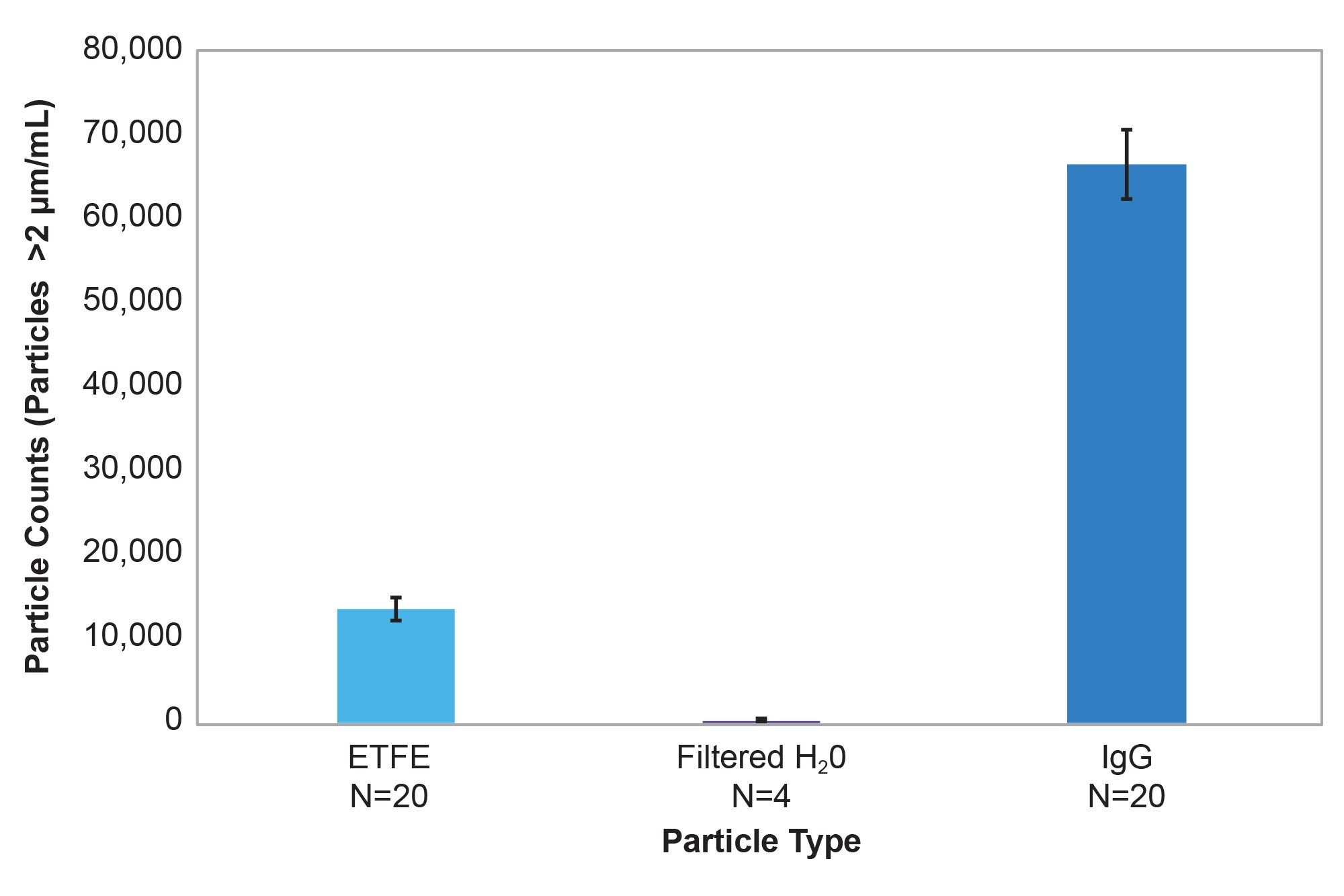
Subvisible particle measurements using 25 μL of highly disperse particle populations of agitated Int-mAb ( n = 20) aggregates, ETFE (n = 20) and water control (n = 4) were performed with BMI (Figure 1). The average of the 20 replicates of Int-mAb IgG samples were measured at 66,632 particles/mL with a CV of 6.2%, while the average of particles measured for ETFE was 13,580 with a CV of 9.7%. The water control had low counts, (an average of eight particles on the membrane) with particulates in the water control ranging from 1–11 particles per well. Total experiment time to analyze 44 samples was less than 60 minutes using the HORIZON Instrument — 30 seconds to background, 30 seconds to measure the sample, plus pipetting time and sample preparation.
Study 2 — Impact of Volume on IgG Aggregates and ETFE Particle Counts
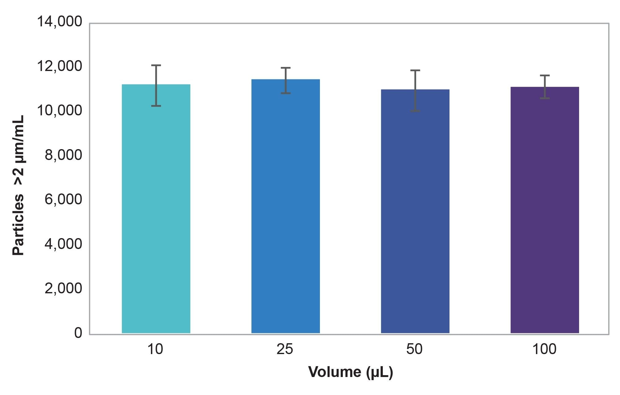
Next, sample size impact on biopharmaceutical subvisible analysis on BMI was tested by measuring several volumes of an ETFE sample on the HORIZON Instrument (Figure 2). Samples measured at volumes of 10 μL, 25 μL, 50 μL, and 100 μL (n = 6 wells for each volume) generated particle counts within 4% of each other for particles greater than 2 μm, proving equivalent counts were obtained when 10 μL and 100 μL of sample were analyzed. The average particle count was 11,121 particles/ mL with CVs ranging from 5.0% to 8.1%, demonstrating system robustness. Thus, precise particle counts can be obtained using 10 μL of sample. But when sample is limited, however, it is recommended using 25 µL for more quantitative work.
Conclusion
BMI analysis of ETFE and IgG aggregates using as little as 25 μL of sample precisely measured both highly polydisperse and low refractive index contrast biopharmaceutical samples. Also, particle counts obtained with as little as 10 μL of solution were equivalent to counts obtained with 100 μL, as demonstrated for the ETFE solution measured at various volumes. Thus, precise subvisible particle measurements can be generated with as little as 10–25 μL of material using BMI, though 25 μL should be used for quantitative work.
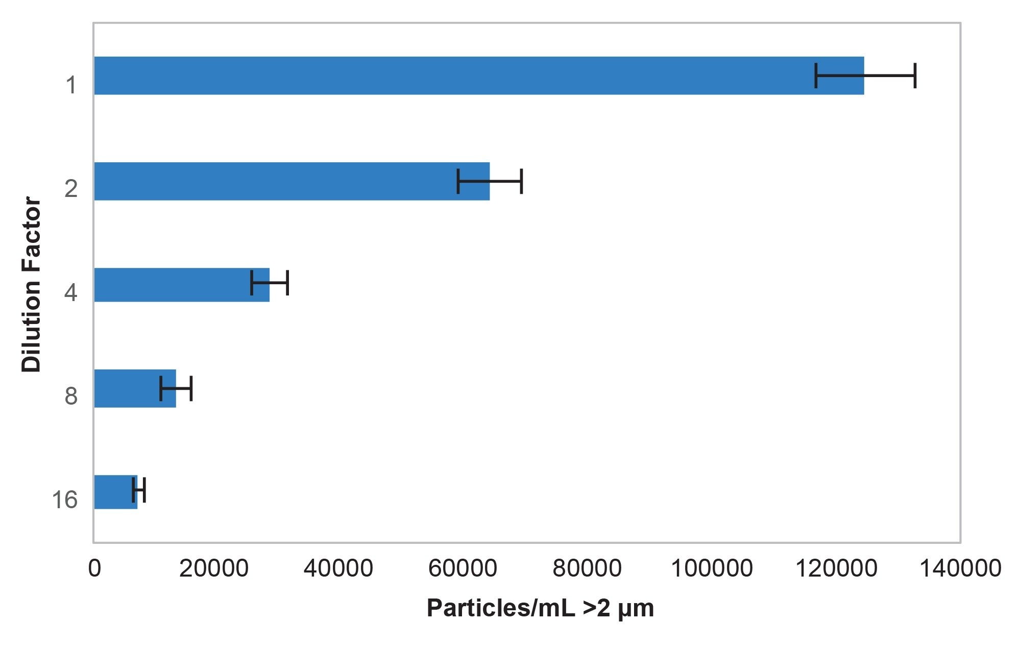
The HORIZON System exhibited linear behavior during serial dilution experiments of Int-mAb. For a serial dilution of two, one would expect an outcome where every dilution factor increase of 2X would result in ½ the measured particle counts. This equates to exponentially decreasing particle counts of the formula y = Ax-1 where A is the number of particles at dilution of 1. Fitting an exponentially decreasing line to this data produces a fit of 125668x-1.058 with an R2 value of 0.9979. This shows that there is less than 6% sampling, inter-instrument, and pipetting error (1.058 vs 1.000) for this formulation.
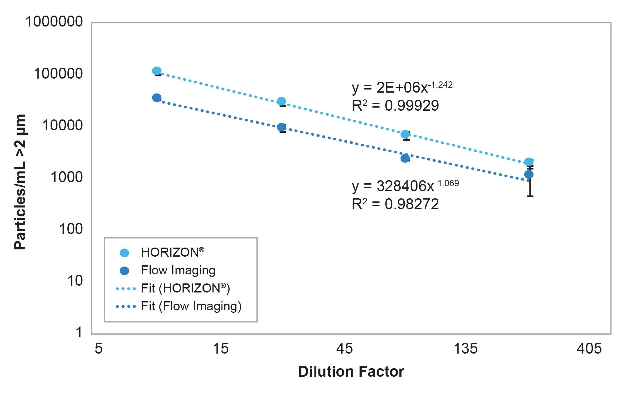
Both FI and BMI were highly linear with respect to each other (R2 fit = 0.9995). However, the FI System required roughly 300 μL per sample with >60% of this volume used for purging the flow cell, while the HORIZON BMI System required 6X less sample. The fact that BMI produced lower CVs, even at lower volumes, can be likely attributed to sensitivity of the BMI technique. The sensitivity is attained because BMI has a greater refractive index contrast between the nearly translucent particles and the surrounding media (see related application note).
The HORIZON System is a fluidics-free subvisible analyzer that is free of the issues that have historically impeded low volume measurements. Powered by BMI, a volume invariant method, the HORIZON Instrument delivers early and robust insights on a formulation’s subvisible particle content by analyzing volumes as low as 25 μL quantitatively and as low as 10 μL for quick, qualitative screens. Ultra-low volume analysis means researchers can now quickly measure if they have an unstable formulation, or get to the right formulation conditions, with just a fraction of the volume to access this key stability parameter as early as candidate selection.
References
Wadhwa, M.; Knezevic, I.; Kang, H. N.; Thorpe, R. Immunogenicity assessment of biotherapeutic products: an overview of assays and their utility. Biologicals 2015, 43 (5), 298–306.
Weiss, W. F.; Young, T. M.; Roberts, C. J. Principles, approaches, and challenges for predicting protein aggregation rates and shelf life. J. Pharm. Sci. 2009, 98 (4), 1246–1277.
Roberts, C. J. Therapeutic protein aggregation: mechanisms, design, and control. Trends Biotechnol. 2014, 32 (7), 372–380.
Roberts, C. J. Non-native protein aggregation kinetics. Biotechnol. Bioeng. 98 (5), 927–938.
Bee, J. S.; et al. Production of particles of therapeutic proteins at the air–water interface during compression/dilation cycles. Soft Matter 2012, 8 (40), 10329–10335.
Mahler, H. C.; Müller, R.; Friess, W.; Delille, A.; Matheus, S. Induction and analysis of aggregates in a liquid IgG1-antibody formulation. Eur. J. Pharm. Biopharm. 2005, 59 (3), 407–417.
Kiese, S.; Papppenberger, A.; Friess, W.; Mahler, H. C. Shaken, not stirred: mechanical stress testing of an IgG1 antibody. J. Pharm. Sci. 2008, 97 (10), 4347–4366.
Ripple, D. C.; Carrier, M. J.; Wayment, J. R. Standards for the optical detection of protein particulates. American Pharmaceutical Review 2012, 14 (5). https://www.nist.gov/publications/standards-optical-detection-protein-particulates
Ripple, D. C.; Montgomery, C. B.; Hu, Z. An interlaboratory comparison of sizing and counting of subvisible particles mimicking protein aggregates. J. Pharm. Sci. 2015, 104 (2), 666–677.
Featured Products
720009618, October 2026


