Lipids play an important role in the energy storage, cellular signaling, and pathophysiology of diseases, such as cancer, neurodegenerative diseases, infections, diabetes, etc. Advances in LC-MS have allowed lipids to be studied with greater sensitivity and specificity. This alleviates the effects of co-eluting compounds and isobaric interference, and allows low abundance lipids to be more readily detected.1
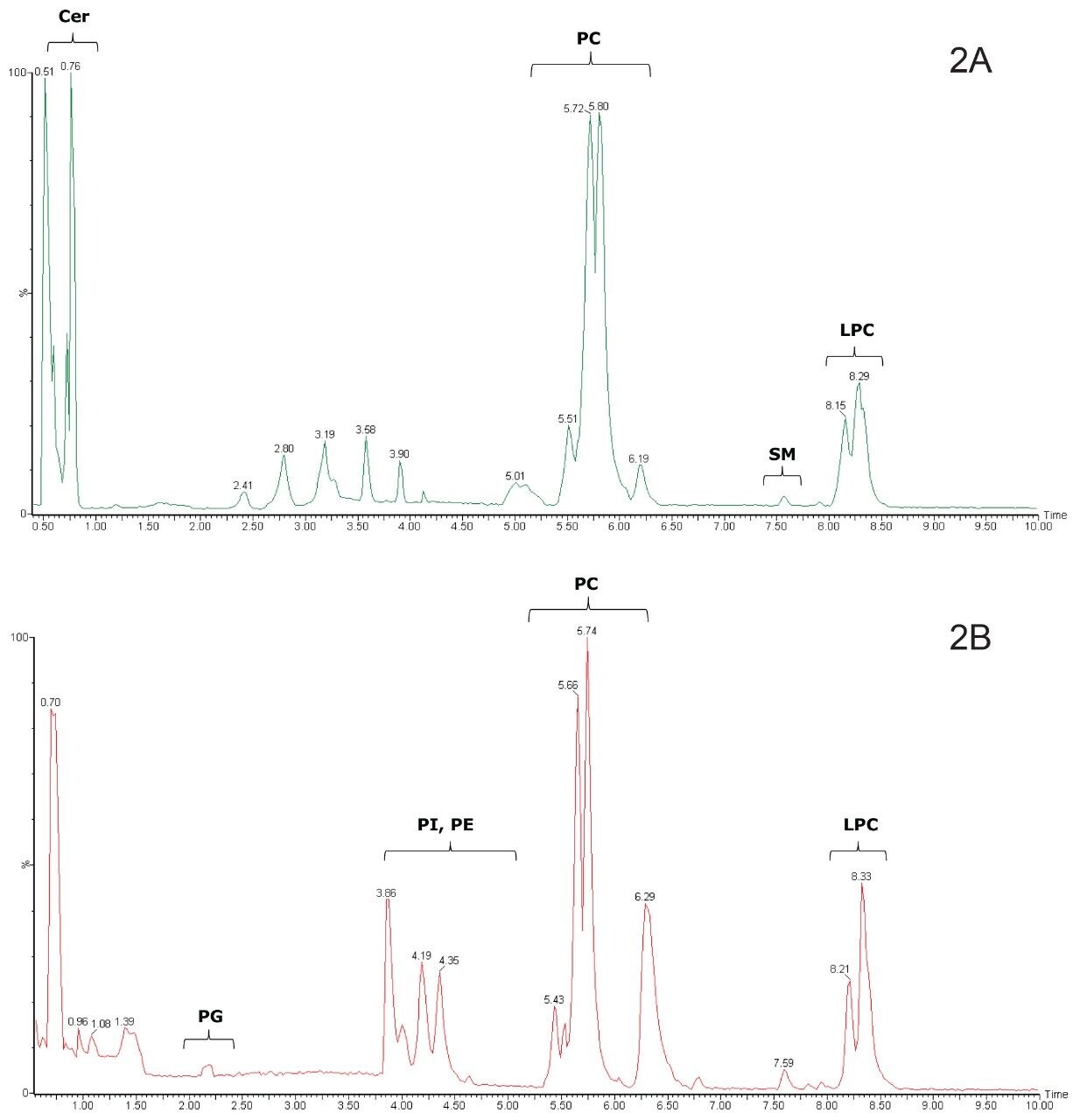
Conventional mass spectrometric analysis of lipids is often performed by direct infusion and reversed-phase or normal-phase HPLC.2-4 However, both normal-phase and reversed-phase methods have certain disadvantages. Although reversed-phase-based separation methods provide good separation of lipids, the complete separation of lipids by class has not been shown.4,5 This is due to the fact that the mechanism of action in reversed-phase chromatography of lipids is based on their lipophilicity, which is governed by the carbon chain length and the number of double bonds.6 As a consequence, co-elution of lipids belonging to different classes in reversed-phase separations is quite common. Normal-phase methods typically allow separation and characterization of different lipid classes, but their lengthy elution and equilibration times make them very time consuming. Moreover the mobile phases typically used for normal-phase – such as chloroform or hexane – lack compatibility with UPLC systems, are difficult to handle due to their volatility and toxicity, and prove challenging for ionization and introduction into a mass spectrometer.7
Although HILIC is considered a variant of normal-phase chromatography, a reversed-phase solvent system (organic-aqueous) can be used to avoid these potential issues. Indeed, due to the highly organic mobile phase (> 80% acetonitrile) used in HILIC, electrospray ionization (ESI) may be improved through more efficient mobile phase desolvation and compound ionization. Performing HILIC using 1.7 μm unbonded BEH particles provides all the acknowledged advantages of UPLC – faster methods, with enhanced chromatographic resolution, sensitivity, and reproducibility of separation.
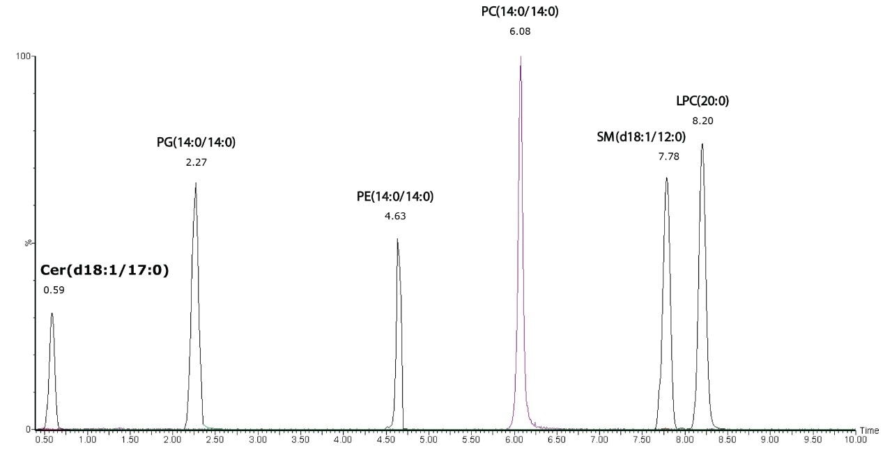
This application note describes a HILIC-based UPLC method that provides better separation of polar lipid classes than existing reversed-phase-based methods, and is faster than previous normal-phase HPLC methods.