
Characterization of Endogenous Ligands in Membrane Proteins Using Dynamic Field Declustering (DFD) on the Cyclic™ IMS P20 Mass Spectrometer
Dale Cooper-Shepherd, Isabella Jones
Waters Corporation, Wilmslow, United Kingdom
Published on September 04, 2026

Abstract
Membrane proteins consitute up to 60% of all approved therapeutic targets and, as such, are important systems to characterize in both therapeutic and biomedical research. However, studying these species by native mass spectrometry remains challenging, as ionization typically requires solubilizing detergents, which can hinder mass measurement. Furthermore, detailed characterization of membrane protein interactions with ligands and other biomolecules can require precise control of ion activation and structural interrogation capabilities. This application note highlights the implementation of Dynamic Field Declustering (DFD), a novel technology on the Cyclic IMS P20 Mass Spectrometer, which effectively removes detergent molecules from ionized proteins to enable accurate mass measurement. For the well-characterized protein bacteriorhodopsin, DFD is shown to completely strip the detergent micelle to reveal high-quality protein as well as endogenous lipid-bound signals. The unique geometry of the Cyclic IMS P20 Mass Spectrometer enabled pseudo-MS4 analysis of a bound lipid allowing its selective dissociation, isolation and fragment-level characterization for unprecedented confidence in endogenous binder assignment.
Benefits
- Enable high confidence assignment of protein signals with DFD
- Confirm endogenous binders from challenging membrane protein samples
- Increase confidence in endogenous binder identification through advanced multi-stage analysis
Introduction
Native mass spectrometry is established as a key tool to characterize protein-ligand interactions.1 The facile nature of measurement through mass-shift detection makes the workflow attractive for drug screening assays and affinity characterization.1,2 Furthermore, catch-and-release type assays enable the detection and characterization of unknown binders directly from biological matrices or endogenous environments.3,4
Membrane proteins represent approximately 30% of the proteome and around 60% of all drug targets and as such are key molecules in drug development as well as biomedical research.5 They remain challenging to study by native mass spectrometry, as they require solubilizing detergents or membrane mimetics to preserve their native structure, which can substantially interfere with mass measurement. To enable effective and accurate mass measurement, it is necessary to remove such solubilizing agents from the protein in the gas phase post-ionization to reveal the ‘naked’ protein. This is usually accomplished using collisional activation after the source region or within a gas cell usually employed for collision-induced dissociation (CID). Modern mass spectrometry (MS) systems, however, often lack the voltage range for complete detergent removal. Furthermore, careful control of this activation step is essential to effectively remove the solubilizing environment while preserving the non-covalent interactions of interest.
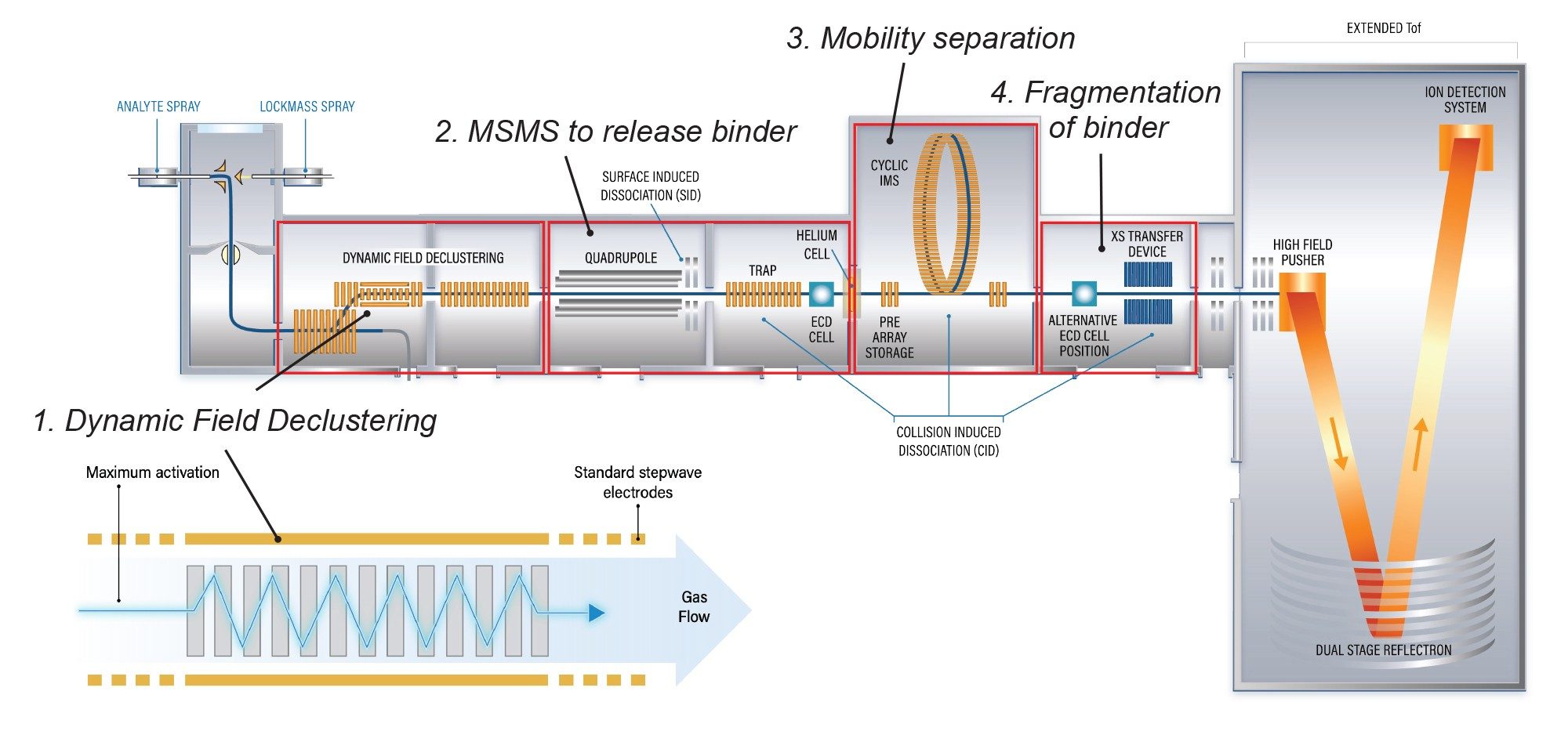
Dynamic Field Declustering (DFD), available on the Cyclic IMS P20 Mass Spectrometer, removes unwanted bound species from proteins enabling highly accurate mass measurement and confident protein characterization. Through sustained activation in an oscillating radio-frequency (RF) field, the interactions between proteins and unwanted residual solvent and other adducts are disrupted, removing them from the protein. In the case of membrane proteins, DFD has been shown to effectively liberate them from their detergent micelle to reveal well resolved protein signals yielding accurate masses. DFD has also been shown to allow retention of binders, e.g. drugs and endogenous lipids.6
The positioning of the DFD device within the Cyclic IMS P20 Mass Spectrometer is central to its functionality. Located in the StepWave™ Ion Guide, DFD enables complete removal of micelles prior to quadrupole mass selection. This allows tandem-MS interrogation of the protein and/or its bound forms by true MS/MS.
This capability was previously demonstrated using prototype instrumentation to characterize a membrane protein-drug complex resulting from a multiplexed small molecule screening campaign.6 In the study, a pseudo-MS4 workflow was employed in which the protein-drug complex was first released from the micelle using DFD, then isolated using the quadrupole. The bound ligand (the drug) was then collisionally released from the complex, subsequently separated from the protein ions by Cyclic ion mobility and, finally, dissociated by CID post-IMS for fragment-level characterization and structural confirmation. By combining efficient micelle removal, selective ion isolation, Cyclic ion mobility separation, and downstream fragmentation, this prototype instrument configuration, now implemented in the Cyclic IMS P20 Mass Spectrometer, enabled confident identification and structural confirmation of the drug candidate binder from the screening campaign.6
In this application note, the pseudo-MS4 workflow is applied on the Cyclic IMS P20 Mass Spectrometer to enable confident characterization of lipid-bound membrane protein complexes. A model system is employed; Bacteriorhodopsin (bR), a membrane-bound, light-driven proton pump from the archaeon Halobacterium salinarum which is often used as a model for G-protein coupled receptors (GPCRs). DFD effectively removes the detergent micelle to reveal well resolved protein signals. Spectral deconvolution shows that signals consistent with bound lipids are observed directly in the mass spectrum. These lipid-bound ions are then interrogated using the pseudo-MS4 workflow, enabling accurate assignment of the lipid species not just from its molecular mass but from its constituent fragments.
Experimental
Sample Preparation
bR in purple membrane isolated from Halobacterium salinarum (Sigma-Aldrich, B0184) was prepared for native analysis by re-suspension in 40 mM (2 x CMC) β-D-glucopyranoside (β-OG) detergent (Sigma-Aldrich) and 200 mM ammonium acetate followed by minimal vortexing. The resulting purple solution was clarified by centrifugation at 10,000×g for 5 minutes and finally buffer exchanged with a BioRad Micro Bio-Spin P-6 gel filtration column.7 The final sample concentration is estimated as 1–5 µM bR monomer.
The sample was electrosprayed from borosilicate glass nanocapillaries (TIP2TW1, World Precision Instruments, FL, USA). Nanocapillaries were mounted onto the NanoLockspray II Ionization Source using the static nanospray adaptor kit (p/n: 186006331). Electrospray voltage was applied to the solution using a piece of platinum wire inserted into the capillary.
Method Conditions
|
MS system: |
Waters Cyclic IMS P20 Mass Spectrometer |
|
Mode: |
Sensitivity |
|
Mass range: |
50–8000 m/z |
|
Polarity: |
Positive |
|
Scan time: |
1 s |
|
Cone voltage: |
50 V |
|
Source temperature: |
100 °C |
|
Capillary voltage: |
1.2 kV |
|
Trap collision voltage (pseudo-MS4): |
100 V |
|
Transfer collision voltage (pseudo-MS4): |
30 V |
|
DFD frequency: |
60 kHz |
|
DFD amplitude: |
175 Vpp |
|
Mobility mode: |
Single Pass |
|
Mobility T-wave amplitude: |
22 V |
|
Mobility T-wave velocity: |
375 ms-1 |
|
Injection time: |
10 ms |
|
Inject array wave amplitude: |
5 V |
|
Inject array offset: |
54 V |
|
Separate time: |
10 ms |
|
Separate array offset: |
55 V |
|
Racetrack bias: |
55 V |
Results and Discussion
Firstly, bR in β-OG was electrosprayed under native conditions. The mass spectrum displayed a series of high intensity signals consistent with detergent clusters (Figure 2A). No protein signals were detected. Upon activating DFD, several high-quality, protein-related signals were revealed (Figure 2B). Deconvolution using the BayesSpray algorithm in waters_connect™ Software yielded an intact average mass for bR of 27049.9 Da, a mass error of 4 ppm versus the theoretical protein sequence mass of 27050.0 Da (Figure 2C). The deconvoluted spectrum also showed signals of 27951.1 Da, 28852.4 Da, 29753.6 Da, mass differences of +901, corresponding to sequential binding of up to three copies of the archaeal lipid PGP-ME (Figure 2C, red circles). Also observed were masses 28269.6 and 29490.0 Da, corresponding to binding of one and two copies of S-TGA-1, respectively, with sequential mass shifts of 1220 Da (Figure 2C, blue squares). Mixed complexes of bR bound to PGP-ME and S-TGA-1 were also observed with masses of 29170.6 and 30072,0 Da, corresponding to bR:PGP-ME:S-TGA-1 ratios of 1:1:1 and 1:2:1, respectively.
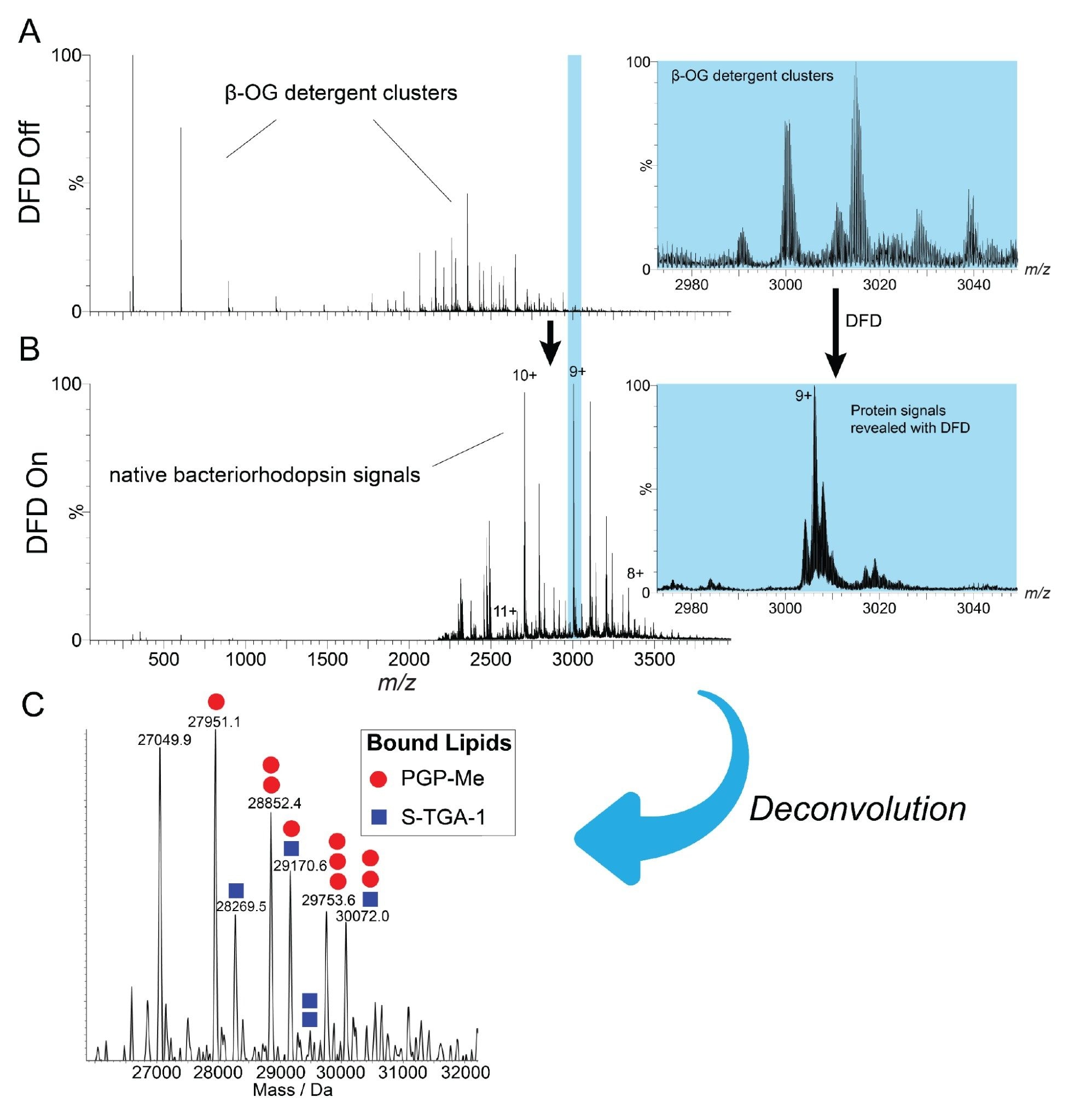
The detection of binding at the intact complex level by native MS does not always provide sufficient confidence for binder identification, especially when analyzing proteins from more complex endogenous environments. To further characterize the bound lipids, a pseudo-MS4 experiment was performed. Firstly, the detergent micelle was stripped using DFD to reveal the lipid-bound protein signals (Figure 2). Secondly, the quadrupole mass filter was used to isolate the ion population corresponding to [bR+PGP-ME]9+. Next, this population was activated in the trap collision cell to release the PGP-ME lipid from the complex.
The resulting spectrum (Figure 3A) shows low-mass lipid derived ions and residual [bR+PGP-ME]9+, [bR]9+ and [bR]8+. The high abundance of [bR]9+ indicates that the lipid primarily dissociates through a neutral loss pathway, but the presence of free lipid [M+H]+ signals and low levels of [bR]8+ indicate that the charged lipid loss pathway is also significant.
Following dissociation, the resulting product ions and remaining precursors are separated using Cyclic ion mobility before undergoing CID in the post-mobility transfer collision cell. This final step produces mobility-aligned product ions, where the fragments appear at the same arrival time as their precursor.
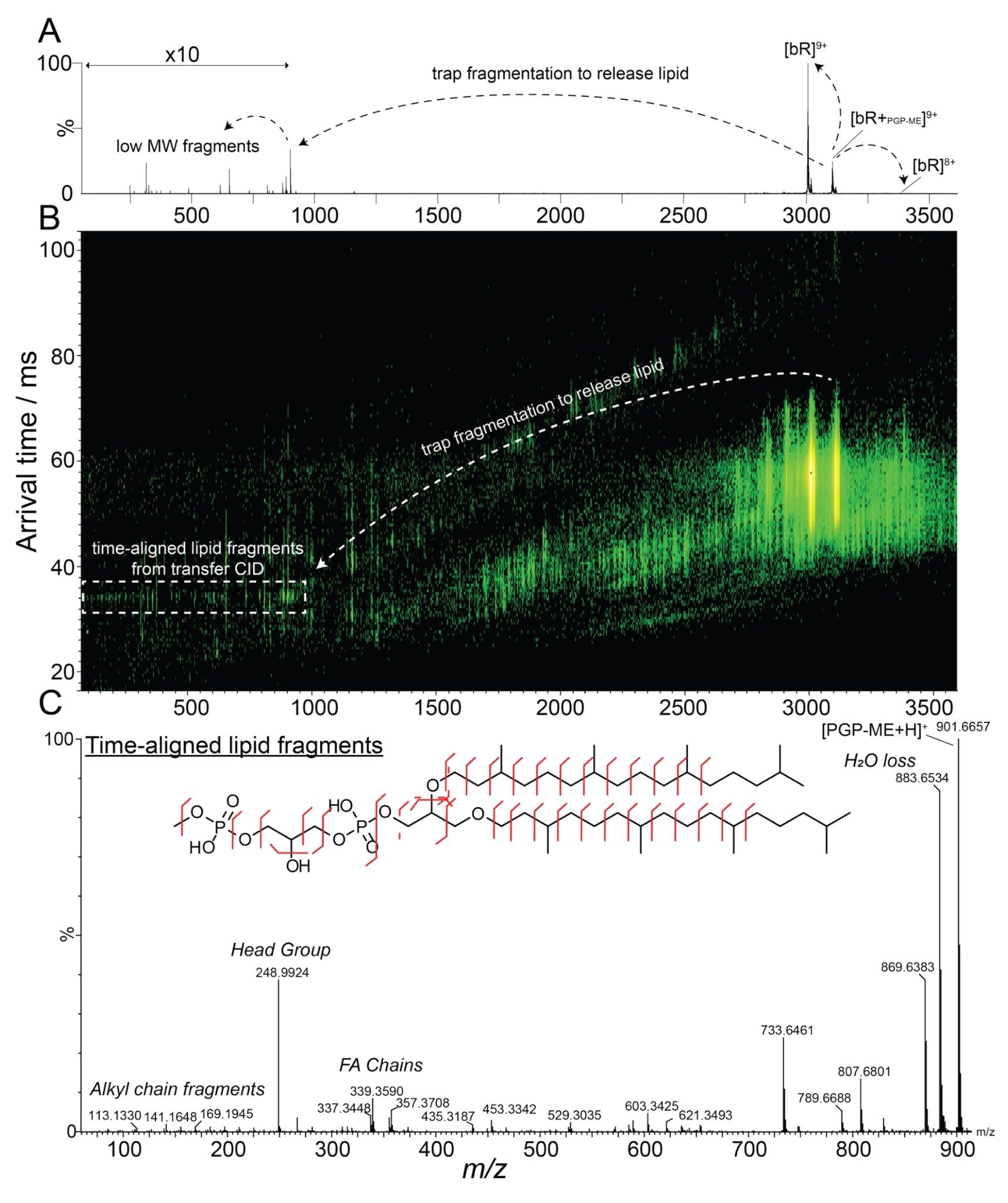
Upon inspection of the low m/z region of the mass spectrum (Figure 3A), a number of signals are observed including PGP-ME at 901.6 m/z and others at 653 and 315 m/z. In the mobility dimension, it can be seen that the protein and low MW species are separated from each other (Figure 3B). Once time-alignment is performed relative to the 901.6 m/z PGP-ME precursor, it is found that the 653 and 315 m/z ions are in fact interfering fragments not originating from the lipid, i.e. they appear at different arrival times to the PGP-ME signal. The resulting time-aligned data (Figure 3C) yield a clean product ion spectrum for the isolated lipid which facilitates high confidence assignment and structural confirmation as PGP-ME. The lipid spectrum displays signals enabling confirmation of the head group (248.99 m/z), the identical fatty acid chains (337.34, 339.36, 355.36 and 357.37 m/z), alkyl chain fragments, as well as other confirmatory ions. All this unprecedented detail on the bound species was obtained directly from a native lipid-bound protein.
Conclusion
The Cyclic IMS P20 Mass Spectrometer provided a powerful workflow for the characterization of membrane protein–lipid interactions by native mass spectrometry. DFD enabled efficient removal of the detergent micelle from bacteriorhodopsin, revealing protein and endogenous lipid-bound species for accurate mass measurement.
The combination of DFD with quadrupole mass selection and Cyclic ion mobility separation enabled a pseudo-MS4 experiment that provided additional structural information for bound lipids, increasing confidence in endogenous binder identification. Together, these capabilities extend the analytical power of native MS and provide new opportunities to study protein–ligand interactions.
References
- Fiorentino, F.; Rotili, D.; Mai, A. Native mass spectrometry-directed drug discovery: Recent advances in investigating protein function and modulation. Drug Discov Today 2023;28:103548. https://doi.org/10.1016/J.DRUDIS.2023.103548.
- Hopper, J.T.S. Mass Spectrometry: Application to Protein-Ligand Interactions. Encyclopedia of Biophysics 2021:1–7. https://doi.org/10.1007/978–3–642–35943–9_216–1.
- El-Hawiet, A.; Shoemaker, G.K.; Daneshfar, R.; Kitova, E.N.; Klassen, J.S. Applications of a Catch and Release Electrospray Ionization Mass Spectrometry Assay for Carbohydrate Library Screening. Anal Chem 2011;84:50–8. https://doi.org/10.1021/AC202760E.
- Gault, J.; et al. Combining native and ‘omics’ mass spectrometry to identify endogenous ligands bound to membrane proteins. Nat Methods 2020;17:505–8. https://doi.org/10.1038/S41592–020–0821–0.
- Errey, J.C.; Fiez-Vandal, C. Production of membrane proteins in industry: The example of GPCRs. Protein Expr Purif 2020;169:105569. https://doi.org/10.1016/J.PEP.2020.105569.
- Sokratous, K.; et al. Enhanced Declustering Enables Native Top-Down Analysis of Membrane Protein Complexes using Ion-Mobility Time-Aligned Fragmentation. J Am Soc Mass Spectrom 2024;35:1891–901. https://doi.org/10.1021/JASMS.4C00190.
- Le, J.; Loo, J.A. Native Top-Down Mass Spectrometry Characterization of Model Integral Membrane Protein Bacteriorhodopsin. J Am Soc Mass Spectrom 2025;36:961–8. https://doi.org/10.1021/jasms.4c00439.
Featured Products
720009573, September 2026