
High-Throughput Stability Screening of Antibody Formulations with Dynamic Light Scattering (DLS)
Bretton Fletcher, Mason Fung, Sophia Kenrick
Waters Corporation, United States
Published on August 31, 2026

Abstract
High-concentration biologic formulations present unique challenges, including aggregation, viscosity, and reduced shelf life. DLS offers a rapid, non-destructive method to assess size distribution, aggregation propensity, and colloidal stability under native formulation conditions. Using the DynaPro™ Plate Reader, scientists can screen hundreds of conditions in parallel with minimal sample consumption and no labeling. In this application note, DLS provided critical insights into colloidal and conformational stability for high-concentration proteins and monoclonal antibodies (mAbs). The comparison to previously-reported differential scanning calorimetry (DSC) data is also presented.
Introduction
As biopharmaceutical development trends toward patient-centric delivery, formulations increasingly target high concentrations (often ≥100 mg/mL) to enable smaller dose volumes and subcutaneous administration. While desirable clinically, elevated concentration can exacerbate colloidal attractions that promote undesirable artifacts like aggregation, high viscosity, and turbidity and can complicate manufacturing and stability management. To make informed formulation decisions, development teams need the ability to generate data rapidly under formulation-strength conditions with multiple robust, orthogonal methods.
DLS measures the hydrodynamic radius (Rh) of proteins and aggregates in solution, simultaneously providing polydispersity and the ability to detect large particles at low concentration. When paired with static light scattering (SLS), DLS also quantifies colloidal stability via intermolecular interaction parameters. Both the diffusion interaction parameter, kD, measured by DLS and the second virial coefficient, A2, measured by SLS quantify whether molecules tend to attract or repel each other in solution. Net attractive interactions, quantified by kD < 0, are strongly correlated to adverse solution properties, including high viscosity, propensity to form aggregates, and turbidity at high concentrations, with recent work showing that kD analysis in dilute conditions was the only measure to predict developability issues.1,2 Moreover, favorable solution behavior was not inherently associated with compromised pharmacokinetics, even for antibodies with high isoelectric points or positive charge, suggesting that colloidal stability measured with DLS is well-suited for screening protein candidates.
In addition to colloidal stability, conformational stability can be a critical quality attribute (CQA) in formulation development. Indicators of conformational stability include the temperature at the onset of unfolding (Tonset), melting temperature at the midpoint of the unfolding (Tm), and enthalpy of these phase transitions. Differential scanning calorimetry (DSC) provides the gold standard for measuring these transitions, and multiple references provide data for the sequential unfolding of key domains in mAb products.3,4 DLS and SLS can also be applied to these systems to identify thermal transitions and positively identify pure unfolding from unfolding and aggregation. Together, DLS and DSC offer orthogonal insights into colloidal and conformational stability, enabling formulation scientists to select buffers and excipients that minimize aggregation and maintain product quality throughout development and storage.
Experimental
Lysozyme (GoldBio) was prepared from powder at 30, 70, and 100 mg/mL in a solution of 0.1 M glycine and 0.02 % w/v sodium azide at pH 2.5. Lower concentrations were prepared by diluting into glycine solution from 30 mg/mL stock. Trastuzumab was prepared in histidine and succinate buffers at ~20 mg/mL stocks and diluted into buffer for final concentrations. Histidine buffer was composed of 4 mM histidine and 18.4 g/L trehalose. Succinate buffer was composed of 10 mM succinate and 60 g/L sucrose.
All samples were filtered through 0.02 µm Whatman™ Anotop™ syringe tip filters (Cytiva) before loading onto 384-well microplates (Aurora Microplates), with 30 µL of sample loaded into each well, three replicates loaded per sample concentration, and all wells sealed with oil to prevent evaporation. Plates were centrifuged for 1 minue at 100 x g to remove bubbles and calibrated with lot-certified 40 kDa dextran (Waters Corporation) prepared at concentrations of 2 mg/mL to 10 mg/mL in ddH2O to enable SLS measurements of molar mass.
Measurements were performed with a DynaPro Plate Reader using DYNAMICS™ Software. Data was initially collected at 25 °C before temperature ramps began. Temperature ramps were performed from 25 °C to 85 °C with a difference in temperature of approximately 1 °C between subsequent measurements of each well.
Results and Discussion
High Concentration Lysozyme
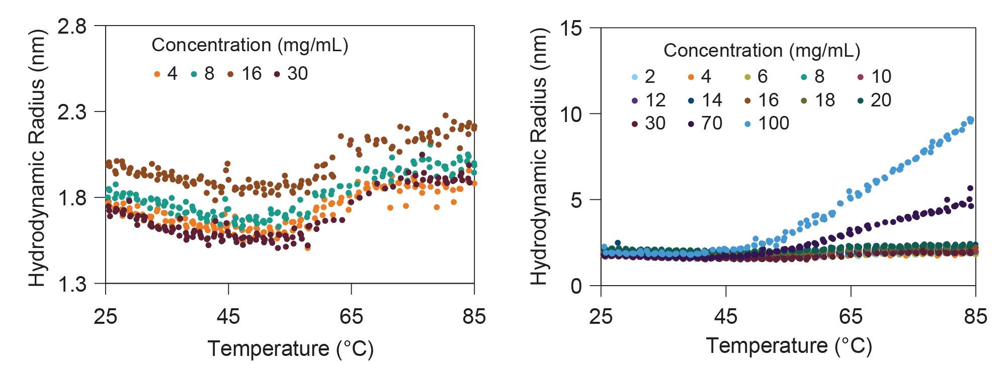
The thermal stability of lysozyme was assessed as a function of protein concentration via DLS measurements of melting and aggregation temperatures. Figure 2 shows representative unfolding curves for lysozyme, with hydrodynamic radius plotted as a function of DLS temperature at protein concentrations of 4, 8, 16, and 30 mg/mL. The hydrodynamic size changed by ~ 0.3 nm across the temperature range from 25 °C to 85 °C, consistent with protein unfolding without aggregation. The onset of unfolding occurred near 58 °C for all conditions with Tm = (62.1 ± 0.7) °C.
In contrast, Tonset for lysozyme prepared at 70 mg/mL and 100 mg/mL was much lower (42.3 °C and 40.9 °C, respectively). Similarly, the hydrodynamic size measured at these higher concentrations increased beyond the expected values for pure protein unfolding and continued to increase across the entire temperature range, reaching Rh > 5 nm. Together, these measurements provide clear evidence of aggregation rather than the pure unfolding observed at lower concentrations.
Trastuzumab in Two Buffer Conditions
Previous DSC analysis of trastuzumab in multiple buffer conditions revealed significant differences in conformational stability as a function of buffering agent and excipient.5 In that study, histidine buffer (4 mM histidine, 18.4 g/L trehalose) was found to be highly stabilizing whereas succinate buffer (10 mM succinate, 60 g/L sucrose) was found to be the most destabilizing. Thus, these conditions were chosen for further analysis by DLS and SLS. Trastuzumab was arrayed from 1 mg/mL to 20 mg/mL in triplicate under both formulations in a single plate. Measurements at 25 °C were performed to assess colloidal stability, and a temperature ramp was performed to quantify unfolding and aggregation, orthogonal to previously published DSC measurements.
Colloidal Stability at 25 °C
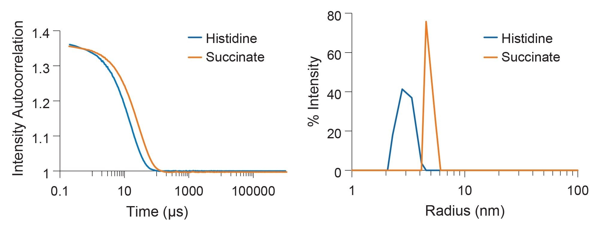
The DLS data at 25 °C indicates that the samples are uniform with hydrodynamic radii around 5 nm, minimal polydispersity, and no other size populations in the solution (Figure 3). Similarly, molar mass measured simultaneously via SLS is consistent with antibody monomer around 150 kDa.
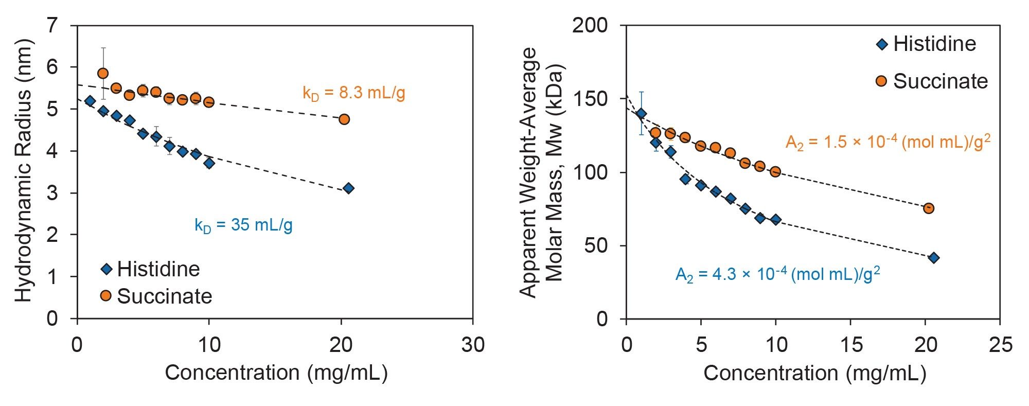
In both formulation buffers, the measured Rh decreased with increasing protein concentration, indicative of repulsive intermolecular interactions. As shown in Figure 4, Rh decreased more steeply in histidine buffer (blue) compared to succinate buffer (orange), suggesting that the histidine buffer promotes stronger repulsive interactions. The strength of these repulsive interactions is quantified by the diffusion interaction parameter, kD, where higher positive values correspond to stronger repulsive interactions. Accordingly, the kD measured for trastuzumab is around 35 mL/g in the histidine buffer and 8 mL/g in the succinate buffer, in agreement with the stabilizing effect of histidine buffer observed in via DSC.5 Similar information can be derived via the second virial coefficient A2 calculated from simultaneous SLS measurements. Figure 4 shows weight-averaged molar mass (Mw) as a function of protein concentration where a clear negative trend in Mw is observed in both buffers due to repulsive intermolecular interactions. In agreement with kD analysis via DLS and with the previous DSC results, A2 analysis for trastuzumab in these two buffer conditions indicates that histidine buffer promotes stronger repulsive interactions, indicative of increased colloidal stability compared to succinate buffer.
Conformational Stability
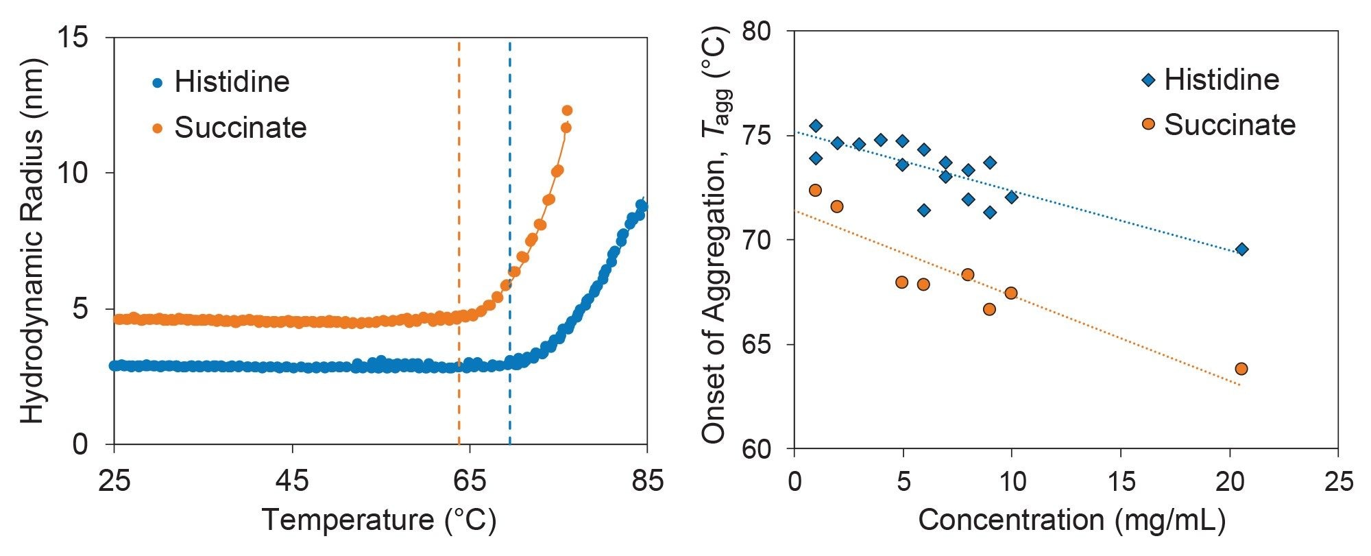
The same samples were further characterized for conformational stability by measuring the change in hydrodynamic size from 25 °C to 85 °C. In agreement with previous DSC data, DLS revealed trastuzumab unfolded at lower temperatures in succinate buffer compared to histidine buffer. Figure 5 shows Rh as a function of temperature for trastuzumab prepared at 20 mg/mL in both buffers. In this case, the histidine buffer appears to promote conformational stability in addition to colloidal stability, as the onset of aggregation occurs around 70 °C, compared to 64 °C in succinate buffer. These measurements are in good agreement with the orthogonal DSC data where a 3 °C difference in denaturation midpoint temperature (Tmax) was observed between the two formulations for the unfolding of the CH2 domain (Tmax = 70.76 °C and 67.71 °C for histidine and succinate buffers, respectively5). Differences in exact temperatures between the two techniques could be due to differences in temperature ramp rates, as explored in a previous application note.6
The high-throughput capabilities of the DynaPro Plate Reader mean that the thermal unfolding can be probed across multiple concentrations of the antibody simultaneously. In this case, lowering the concentration of antibody increases the temperature at which aggregation occurs (Figure 5). This behavior can be expected since lower concentrations decrease the probability with which the unfolding protein molecules may interact. Nevertheless, the onset of aggregation in histidine buffer occurred at consistently higher temperatures as compared to succinate buffer, with differences in Tonset ranging from 2 °C to 6 °C.
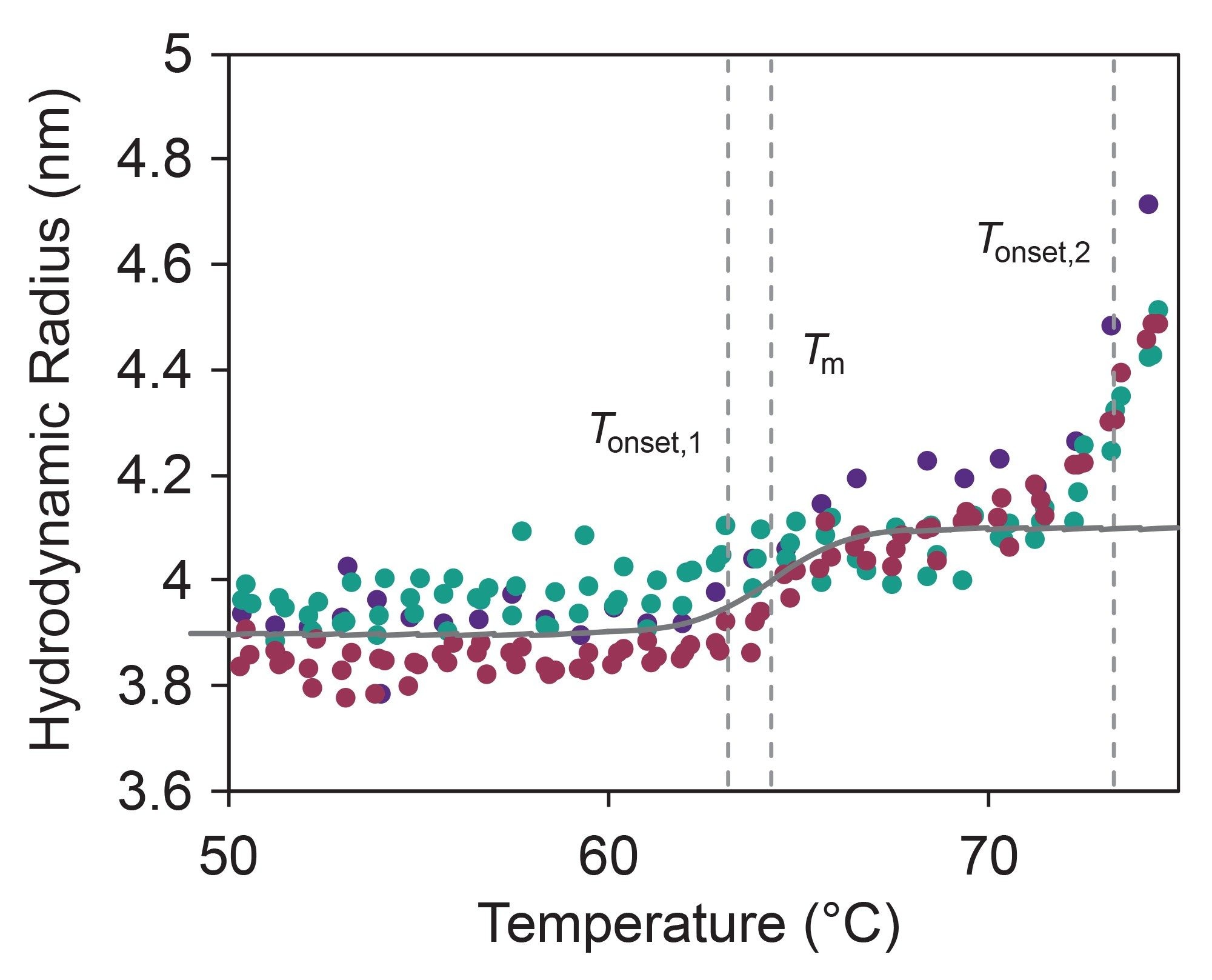
For lower concentrations of trastuzumab in histidine buffer, the DLS data revealed more than just the onset of aggregation. Zooming in between 60 °C and 70 °C, a small but significant size increase is observed from 3.9 nm to 4.1 nm (Figure 6), with no corresponding increase in molar mass. This change in size is indicative of a first unfolding event occurring at 63 °C (Tonset,1), with Tm = 64 °C, before the onset of aggregation at 73 °C. These complement the orthogonal DSC measurements, which found the onset of unfolding Tonset = 65.0 °C, with Tm,1 = 70.76 °C and Tm,2 = 82.66 °C, corresponding to the CH2 and Fab/CH3 unfolding events, respectively.5
Conclusion
In this application note, DLS combined with SLS provided a powerful approach for assessing protein stability under formulation-relevant conditions. High-throughput DLS and SLS measurements with the DynaPro Plate Reader enable rapid, multiplexed characterization of protein size, aggregation, intermolecular interactions, and conformational stability. Together, these data deliver direct insight into formulation stability to support patient-centric delivery routes.
The lysozyme and trastuzumab case studies illustrate how formulation composition and protein concentration jointly influence conformational and colloidal stability and how these effects can be resolved through careful analysis of size, interaction parameters, and temperature-dependent behavior. Comprehensive, high-throughput light scattering measurements can guide formulation selection and risk mitigation early in development, complementing other techniques like DSC by providing an orthogonal assessment of stability with additional insight into size and molar mass. By integrating DLS and SLS measurements with the DynaPro Plate Reader formulation, scientists can rapidly screen conditions, identify stabilizing excipients, and build a more complete understanding of protein behavior across concentration and temperature space.
References
- Kingsbury, A.; et al A single molecular descriptor to predict solution behavior of therapeutic antibodies. Science Advances, 2020.
- Menzen, T.; Friess, W. "Temperature-Ramped Studies on the Aggregation, Unfolding, and Interaction of a Therapeutic Monoclonal Antibody," Journal of Pharmaceutical Sciences, pp. 445-455, 2014.
- Seelig, J.; Seelig, A. "Molecular understanding of calorimetric protein unfolding experiments.," Biophysical Reports, 2022.
- Privalov, P.; Khechinashvili, N. "A thermodynamic approach to the problem of stabilization of globular protein structure: a calorimetric study," Journal of Molecular Biology, pp. 665-684, 1974.
- TA Instruments, "Rapid Thermal Stability Screening and Selection of Monoclonal Antibdy Drug Products," [Online]. Available: https://www.tainstruments.com/pdf/literature/MC178.pdf.
- Waters Corporation, "Influence of heating rates on the thermostability characterization of antibodies by DLS and SLS," [Online]. Available: https://www.wyatt.com/library/application-notes/an4003-influence-of-heating-rates-on-the-thermostability-characterization-of-antibodies-by-dls-and-sls.html.
Featured Products
720009561, August 2026


