
Forced Degradation Method Development for Oncologic Drug Daraxonrasib Using a Systematic Approach and MaxPeak Premier Columns
Kenneth Berthelette, Chris Collins, Thomas H Walter, Paul Rainville
Waters Corporation, United States
Published on October 28, 2026

Abstract
A forced degradation study of the oncologic drug daraxonrasib was performed with the resulting degradation samples combined and a single analytical method developed. To facilitate the development of the method, a structured approach was employed along with inert hardware chromatographic columns. A suitable method was determined after optimization to resolve a critical pair using the ACQUITY™ Premier BEH™ C18 Column with a high pH modified mobile phase and acetonitrile strong solvent.
Benefits
- Baseline resolution of all degradants achieved using systematic method development
- Demonstration of optimizing a method to improve resolution as well as decrease cycle time
Introduction
Drug development encompasses a complex set of workflows ranging from ADME (adsorption, distribution, metabolism, excretion) studies to stability testing, and monitoring impurities. One enabling factor that can be applied to many of these workflows is using a single analytical method to separate the active pharmaceutical ingredient (API), degradants, and process impurities. Developing such a method would allow subsequent testing to be standardized, reducing the need for future work and aligning multiple labs on the optimal conditions. However, developing a method can be challenging and often relies on expert analysts. Implementing structured method development protocols allows less experienced users to rapidly develop methods, which can then be optimized and implemented across different workflows.
One example of a structured method development procedure is the systematic screening protocol, which is a tiered approach for new method generation.1 This protocol has been used previously to develop methods for a variety of samples such as drug impurity mixtures, kinase inhibitors, radiotherapeutic precursors, and many others.1-4 The protocol starts with setting clear and concise separation criteria so that method development has a definitive endpoint. Next is column and mobile phase screening followed by optimization if it is warranted. Getting the most out of this protocol relies on selecting appropriate column stationary phases that maximize the range of selectivity while also offering reliable and reproducible performance for the lifetime of the method. To highlight the use of this protocol, the oncologic drug daraxonrasib was subjected to forced degradation conditions and a method was developed to separate the resulting mixture.
Daraxonrasib was approved by the FDA in August 2026 for the treatment of metastatic pancreatic cancer. This compound acts as a molecular glue, forming a complex with cyclophilin A and active GTP‑bound RAS, thus blocking RAS signaling and limiting cancer growth.5 Clinical trials showed that daraxonrasib, which is offered as a daily oral tablet, improved median overall survival of patients from approximately six months to over a year.6
Experimental
Sample Description
A stock solution of daraxonrasib was created at 1 mg/mL in 60:40 water:acetonitrile (v/v). Two 1 mL aliquots of the stock solution were removed and placed into separate vials. To each vial, 0.1 mL of the appropriate catalyst was added. For acidic hydrolysis, a 1 N hydrochloric acid solution was added and the vial was placed on a hot plate set to 70 °C for one hour. For basic hydrolysis, a 0.5 N sodium hydroxide solution was added and allowed to sit at room temperature for approximately one minute. The two vials were then combined and transferred to TruView™ pH Control LCMS Certified Maximum Recovery Vials and placed on the system for analysis.
LC Conditions
|
LC system: |
ACQUITY UPLC™ H-Class Plus with Column Manager, 2 Column Manager Aux, PDA Detector, and QDa™ Mass Detector |
|
Detection: |
UV @ 254 nm, Full Scan MS with ESI+ Ionization |
|
Columns: |
ACQUITY Premier BEH C18 Column, 2.1 x 50 mm, 1.7 µm (p/n: 186009452) ACQUITY Premier CSH™ C18 Column, 2.1 x 50 mm, 1.7 µm (p/n: 186009460) ACQUITY Premier CSH Phenyl-Hexyl Column, 2.1 x 50 mm, 1.7 µm (p/n: 186009474) ACQUITY Premier BEH Phenyl Column, 2.1 x 50 mm, 1.7 µm (p/n: 186010336) Atlantis™ Premier BEH C18 AX Column, 2.1 x 50 mm, 1.7 µm (p/n: 186009366) |
|
Vials: |
TruView™ pH Control LCMS Certified Clear Glass Vial, Maximum Recovery Vials (p/n: 186005668CV) |
|
Column temperature: |
30 °C |
|
Sample temperature: |
10 °C |
|
Injection volume: |
1 µL |
|
Flow rate: |
0.4 mL/min |
|
Mobile phase A: |
Water |
|
Mobile phase B: |
Acetonitrile |
|
Mobile phase C: |
Methanol |
|
Mobile phase D1: |
2% formic acid in water (diluted to 0.1% on column) |
|
Mobile phase D2: |
200 mM ammonium hydroxide in water (diluted to 10 mM on column) |
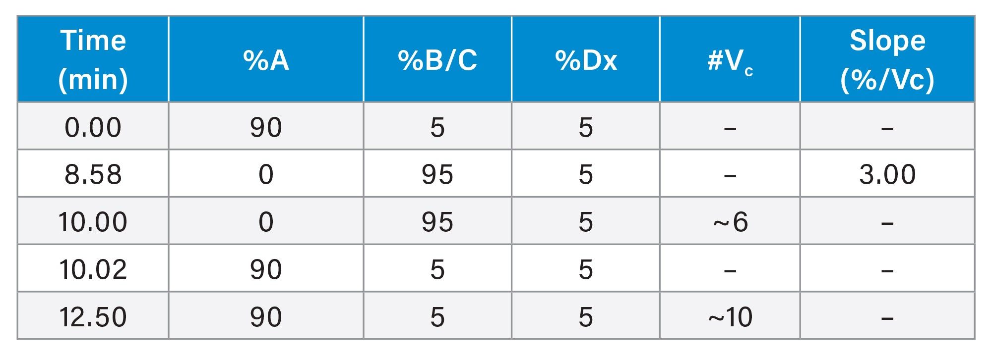
Scouting Gradient Conditions
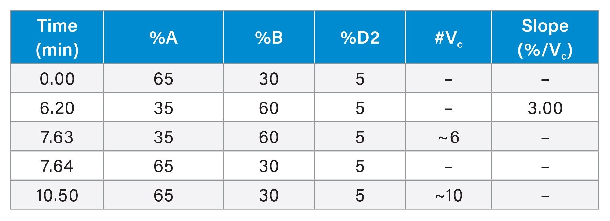
Final Method Conditions
Data Management
|
Chromatography software: |
Empower™ 3 Feature Release 5 (FR5) |
Results and Discussion
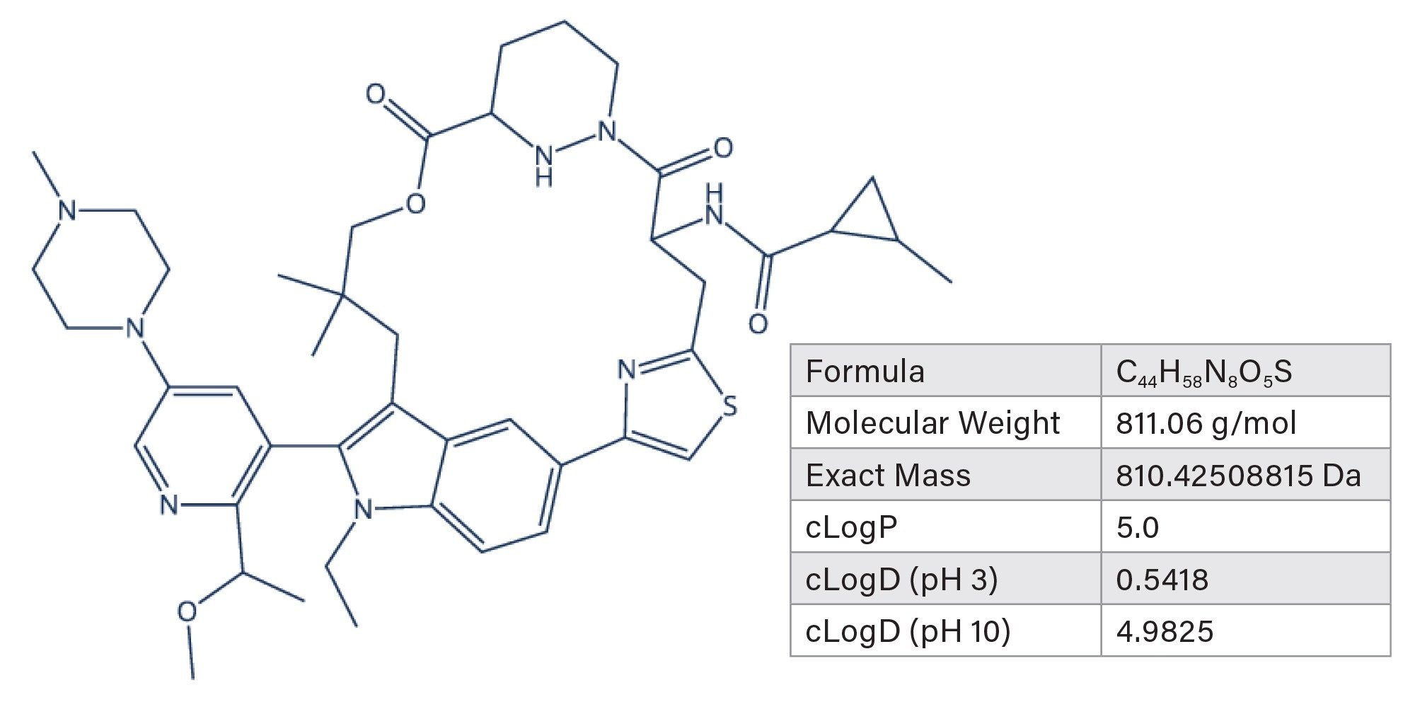
Developing methods is considerably easier when employing a structured protocol. In this work, the systematic screening protocol was used, as it has shown to be a highly effective approach to method development.1-4 The first step of the systematic screening protocol is to define the sample and the separation criteria. This crucial step must be taken prior to any experiments as it can drastically influence the process. In most cases, a sample can be defined with simple descriptors such as the number of analytes present and their chemical properties. However, in some cases, the exact number of analytes in a sample and their chemical properties are not known. This is the case in forced degradation studies. For the analyte that is known, daraxonrasib, Figure 1 shows some of the chemical properties along with the chemical structure. The separation criteria define the end point for the method development process. Common criteria include adequate retention of all analytes, baseline resolution for all peaks, symmetrical peaks, and/or short run times. For the forced degradation of daraxonrasib, the separation needed to achieve baseline resolution of isobaric compounds, as defined by USP resolution values ≥1.5. Additionally, having symmetrical peaks, as defined as USP tailing values between 0.8-1.2 was required. No other criteria were set for this separation.
Once the sample and the separation criteria have been defined, the experimental runs can begin with a determination of which mobile phase pH is better for the analytes. To do this, a hybrid organic/inorganic particle column should be used. Hybrid particles have wider compatible pH ranges compared to their silica particle counterparts, allowing for the same stationary phase to be used at low and high pH.7 For this work, the ACQUITY Premier BEH C18 Column was selected. Figure 2 shows the pH scouting experiments run for the forced degradation sample.
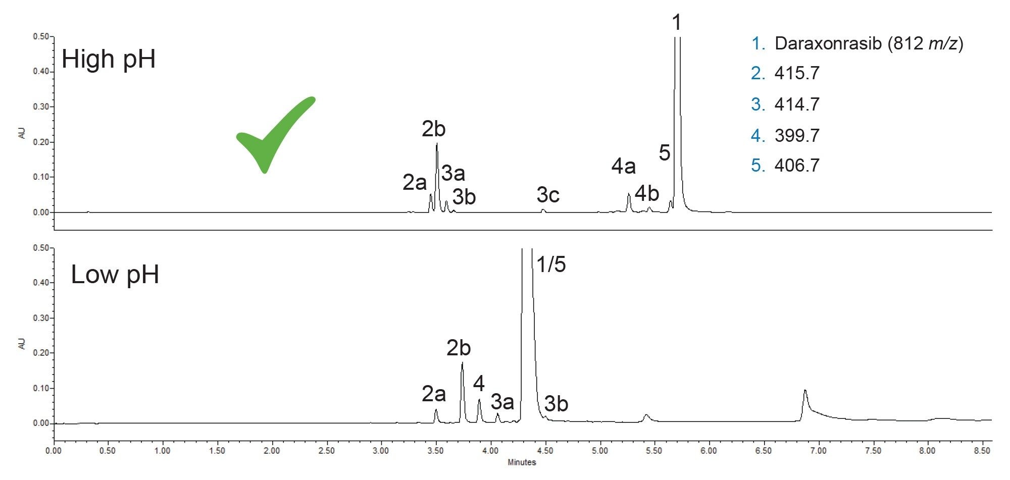
The forced degradation produced a total of eight new peaks, with several having the same mass-to-charge (m/z) ratio. Notably, the m/z ratios of 415.7, 414.7 and 399.7 corresponded to two or three peaks in the resulting chromatograms. Additionally, a component with m/z of 406.7 eluted very close to the daraxonrasib peak (1). Given that this step focuses on the retention of the analytes, high pH is the better choice. While the low pH mobile phase does offer slightly higher retention for components 2b, 3a and 3b, daraxonrasib was considerably better retained and gave a sharper peak at high pH. Consequently, the high pH mobile phase additive was used for all subsequent testing.
The next step in the method development process is the screening of multiple stationary phases, which are chosen to span a range of selectivities while operating at the mobile phase pH selected previously. For this analysis, which was performed with high pH mobile phases, silica-based stationary phases were not selected, as silica hydrolyzes at pH values greater than about,8 leading to column voiding.7 The first column selected was the ACQUITY Premier BEH C18 Column used in the first step. This column provides a solid starting point for many method development activities.
The next stationary phase selected for this work was the charged surface hybrid (CSH) C18, which incorporates a basic functionality in its design,8 leading to some differences in selectivity compared to BEH C18. The next two stationary phases selected were BEH Phenyl and CSH Phenyl-Hexyl. Apart from the presence of a basic functionality in CSH Phenyl-Hexyl, these two stationary phases are similar. The phenyl structure in these bonded phases provides increased retention of aromatic compounds through π-π interactions, especially when methanol is used as the strong solvent. Lastly, the BEH C18 AX stationary phase was selected, as it employs a mid-coverage C18 bonding along with an anion exchange functional group.9 Figure 3 shows the chromatograms for the sample on all five columns tested using methanol as the strong solvent.
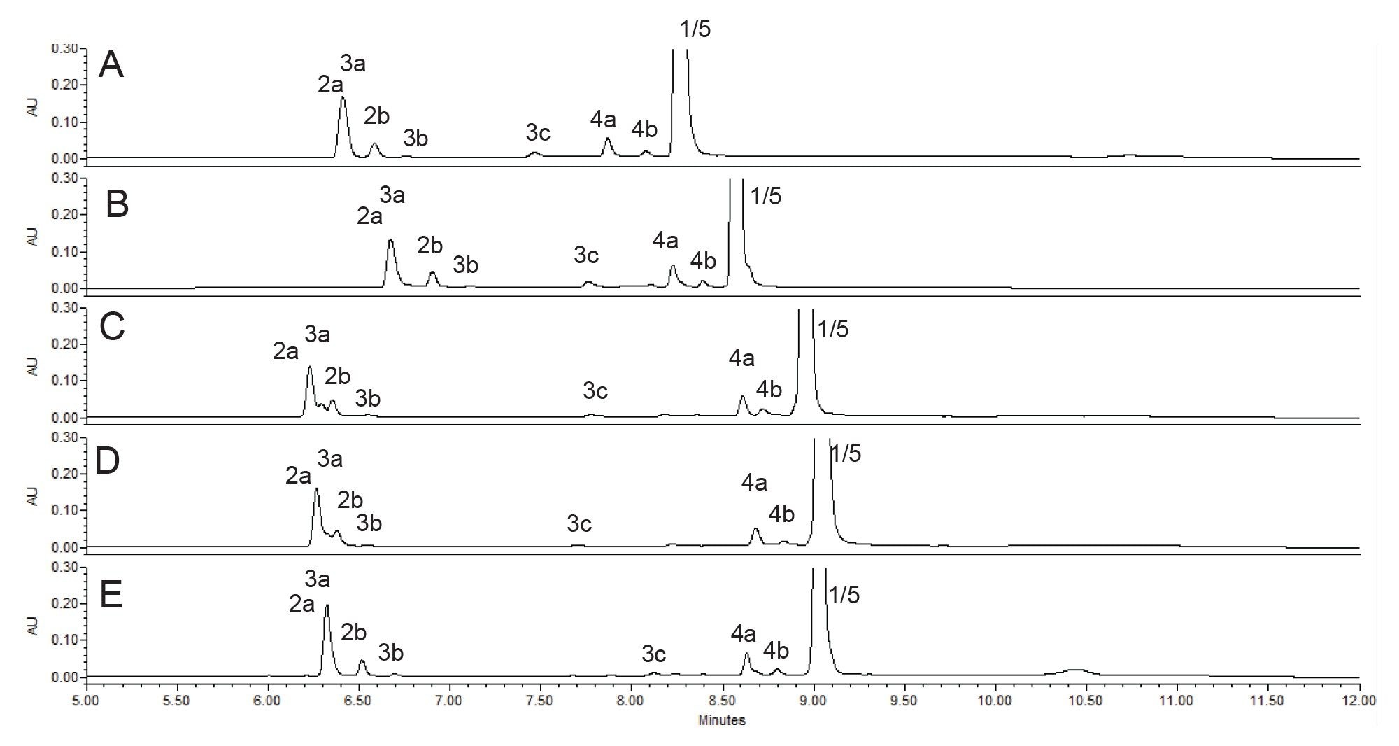
Using methanol as the strong solvent did not yield a separation meeting the method criteria. There was considerable co-elution with all five stationary phases, the most concerning one being the co-elution of component 5 and daraxonrasib (1). Looking at selectivity differences, comparing column A and column C, BEH C18 and BEH Phenyl respectively, there were some interesting differences. First, one of the component 3 peaks co-eluted with the earliest component 2 peak on the C18 stationary phase but was partially separated on the phenyl stationary phase. This was true for both phenyl columns compared to all of the columns which use C18 bonded phases. Another interesting observation is that the CSH C18 Column (Figure 3B) retained all of the components slightly more than the BEH C18 Column (Figure 3A). This was true for the comparison of the phenyl columns as well, which showed the CSH Phenyl-Hexyl Column (Figure 3D) giving slightly higher retention than the BEH Phenyl Column (Figure 3C).
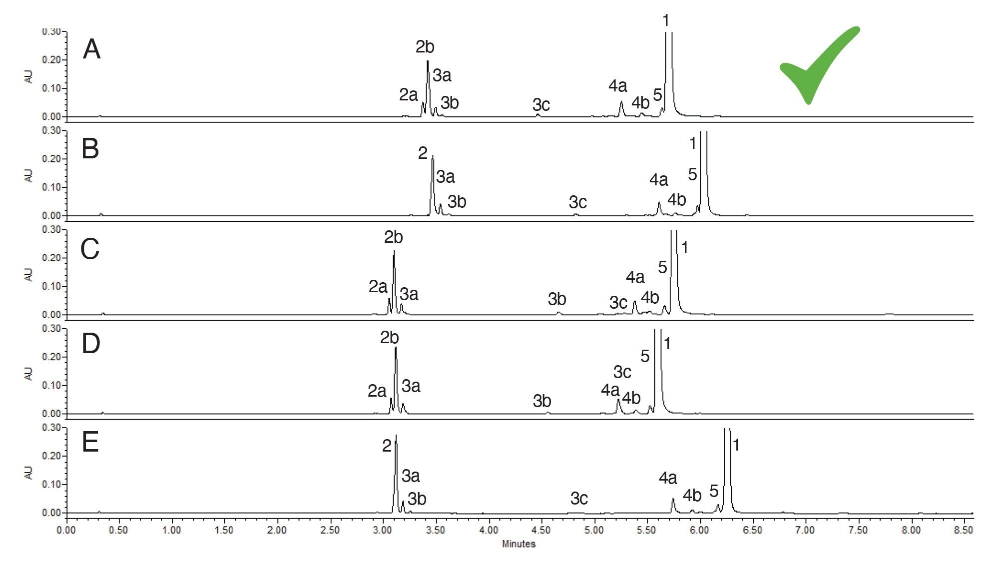
These stationary phases were also tested using acetonitrile as the strong solvent. The results are shown in Figure 4. As with methanol, none of the separations met the method criteria. There was co-elution of at least one set of analytes, component 5 and 1 and some very closely eluting peaks from the component 2 and 3 groups. Of all the separations, the one obtained using the BEH C18 Column with acetonitrile was the best with the highest resolution for the analytes. Method optimization was needed to improve the overall separation quality and meet the separation criteria previously set.
Method optimization steps often include adjustments to the gradient profile, both to the start and end points, but also to the gradient slope. Additionally, changes in column temperature, column configuration, or slight adjustments to the mobile phase pH are possible routes forward during optimization. Adjusting the gradient profile is the fastest parameter to implement and does not result in longer run times as would be the case with using a longer column. For this sample, moving to a 100 mm column would almost certainly provide the separation needed, but at the cost of double the run time. If the separation criteria can be achieved with a shorter run time, that would be preferable. A series of gradient adjustment steps were taken for this sample, and the resulting chromatograms are shown in Figure 5.
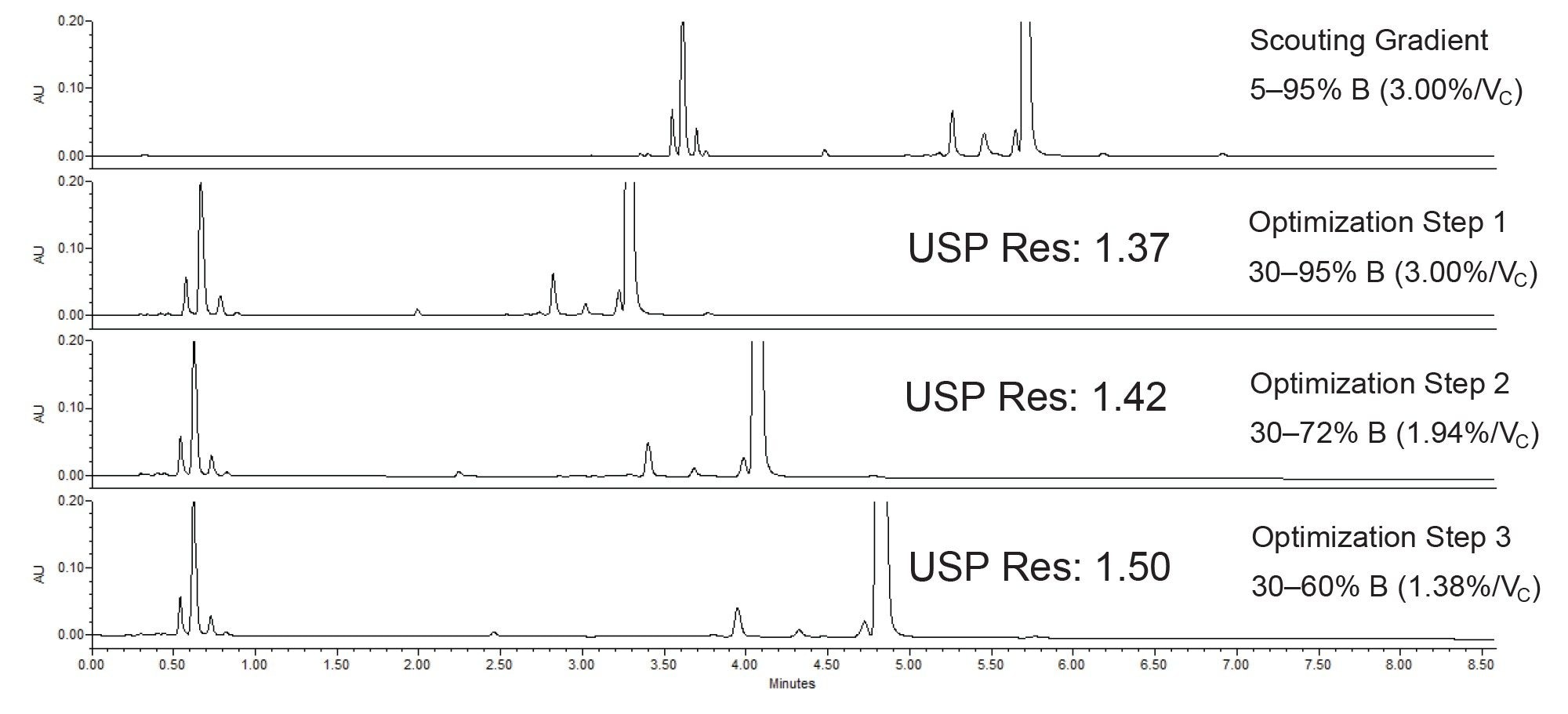
The initial scouting runs used a 5–95% acetonitrile gradient. Examining the chromatogram shown in Figure 4A, the first analyte did not elute until well into the gradient, at approximately 35-40% acetonitrile. To reduce the cycle time, the first gradient adjustment started the method at 30% acetonitrile instead of 5%. The gradient slope of 3.00% per column volume (%/Vc) was maintained to preserve the separation quality. Next, the end point of the gradient was adjusted from 95% to 72%, which not only reduced the cycle time even further, but also decreased the gradient slope from 3.00 to 1.94 %/Vc. As seen in the third chromatogram of Figure 5, the USP resolution increased from 1.37 to 1.42. The last step of optimization reduced the gradient slope even further, down to 1.38 %/Vc which ended up improving the separation enough to meet the criteria set for this analysis. With all criteria met, the separation could now be transferred to QC, scaled up to prep LC in order to purify the degradants and get true identifications, or used for other purposes.
Conclusion
Forced degradation studies are important in drug development and require the creation of a suitable method to separate the degradants. Developing such a method can be challenging, especially for novice chromatographers. To minimize the challenges, a structured method development approach can be employed, leading to faster method development with a traceable decision-making process. To demonstrate how this approach can be used, the oncology drug daraxonrasib was subjected to forced degradation conditions and a suitable LC method was created to separate the intact API from the degradants.
References
- Hong, P.; McConville, P. A Complete Solution to Perform a Systematic Screening Protocol for LC Method Development. Waters White Paper. https://www.waters.com/nextgen/us/en/library/library-details.html?documentid=720005268&t=waters-ACompleteSolutiontoPerformaSystematicScreeningProtocolforLCMethodDevelopment-720005268
- Berthelette, K.; Collins, C.; Haynes, K. Method Development of Proteolysis Targeting Chimera (PROTAC) Compound ARV-825 Forced Degradation Sample Using the Systematic Screening Protocol. Waters Application Note, 720008328, 2024.
- Berthelette, K.; Collins, C.; Kalwood, J.; Haynes, K. Using the Systematic Screening Protocol and MaxPeak Premier Columns to Separate Seven Janus Kinase Inhibitors. Waters Application Note, 720008528, 2024.
- Berthelette, K.; Aiello, M.; Collins, C.; Walter, T.H. Development of a UPLC Method for a Forced Degradation Study of Radioligand Therapy Precursor PSMA-617. Waters Application Note, 720009042, 2025.
- Gierulska, N.; Jankowska, N.; Dabrowska, Z.; Piekarz, J. Skorzewska, M. Efficacy and Safety of Daraxonrasib (RMC-6236) in Previously Treated Metastatic RAS-Mutated Pancreatic Ductal Adenocarcinoma : A Systematic Review. Cancers (2026) 18(19), 3112. https://doi.org/10.3390/cancers18193112
- O’Reilly, M.; et al. Daraxonrasib or Chemotherapy in Previously Treated Metastatic Pancreatic Cancer. New England Journal of Medicine. (2026) 395 (4), 325-337. DOI: 10.1056/NEJMoa2605555
- Wyndham, K.D.; et al. Review of Waters Hybrid Particle Technology Part 2. Ethylene-Bridged [BEH Technology] Hybrids and Their Use in Liquid Chromatography. 720001159, 2004. A Review of Waters Hybrid Particle Technology: Ethylene-Bridged [BEH Technology] Hybrids and Their Use in Liquid Chromatography | Waters
- Iraneta, P.; Wyndham, K.; McCabe, D.; Water, T.H. A Review of Waters Hybrid Particle Technology Part 3. Charged Surface Hybrid (CSH) Technology and Its Use in Liquid Chromatography. Waters White Paper. 720003929. 2011. A Review of Waters Hybrid Particle Technology. Part 3. Charged Surface Hybrid (CSH) Technology and Its Use in Liquid Chromatography | Waters
- Walter, T.H.; et al. Characterization of a Highly Stable Mixed-mode Reversed-phase/Weak Anion-exchange Stationary Phase Based on Hybrid Organic/inorganic Particles. J. Sep. Sci. 44 (2021), 1005-1014 doi: 10.1002/jssc.202001136
Featured Products
720009642, October 2026


