• Applikationsbericht

Cross Validation of an Automated LipidQuan Sample Preparation Procedure

Cross Validation of an Automated LipidQuan Sample Preparation Procedure

  • Nyasha Munjoma
  • Robert Wardle
  • Giorgis Isaac
  • Lee A. Gethings
  • Robert S. Plumb
  • Lisa J. Calton
  • Waters Corporation
Platelet-rich human plasma in tubes

For research use only. Not for use in diagnostic procedures.

This is an Application Brief and does not contain a detailed Experimental section.

Abstract

In this application brief, we cross-validate an automated sample preparation protocol using the Hamilton Microlab STAR Liquid Handling System versus manual preparation for incorporation into the LipidQuan workflow.

Benefits

To evaluate and cross-validate an automated, high throughput sample preparation procedure that could be utilized as a part of the LipidQuan workflow.

Introduction

Lipidomic studies generally require large sample sizes to reliably determine the statistical significance of lipids as potential biomarkers for disease. Furthermore, the availability of large biobanks consisting of thousands of samples represents an important resource, ready-made for interrogation using lipidomic techniques. Access to lipid data from these valuable samples would grant greater insight into the biological mechanisms at the onset and progression of disease.¹

To make the best use of these resources, automated sample preparation strategies are required to miminize cost and experimental variations when extracting lipids from thousands of plasma/serum samples. The most widely used lipid extraction methods are liquid-liquid chloroformmethanol (Folch et al.) and Bligh-Dyer,²which require multiple steps, increasing experimental variability, and complicated automated procedure set up. More recently developed procedures using simple protein precipitation of plasma or serum (e.g., Magali et al., 2014)³ are advantageous in terms of lipid recoveries, safety concerns (i.e., solvents used for the protein crash are less toxic), and the ease of automation for high throughput analyses of large cohorts.

Here we cross-validated a manual protein precipitation procedure against an automated procedure using a Hamilton Microlab STAR Liquid Handling System (Birmingham, UK). LC-MS data were acquired using the LipidQuan solution to provide the identification and quantification of lipids.⁴

Results and Discussion

Automated sample preparation from the LipidQuan protein precipitation step was performed using a Hamilton Microlab STAR to mimic the manual preparation. A simple sample preparation procedure (Magali et al., 2014)³ was adopted for calibrants, QCs, and samples. A 10-point calibration curve and three concentration QC levels of Odd- Chained Lipidomix (Avanti, Alabaster, AL, USA) were spiked directly into commercially available pooled “healthy human plasma” (anticoagulant, K2 EDTA) from Innovative Research (Peary Court, US) at less than 5% v/v of the matrix. The pre-mix standard solution contains lipids from different classes at different concentrations as shown in the CoA. Examples of the various calibration ranges were from 5–650 ng/mL for LPE (17:1) and 1510–1,888,750 ng/mL for PC (17:0/14:1).

Twenty five-microliter (25-μL) plasma aliquots were protein crashed and incubated for two hours at 5 °C using IPA/ACN (1:2, v/v) solution containing a 500x dilution of neat deuterated Ceramide Lipidomix and Splash Lipidomix Mass Spec (Avanti, USA) as internal standards. These standard mixes cover multiple lipid classes and are comprised of heavy (d7–d9) isotopes.

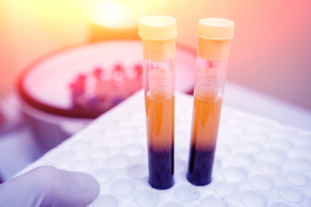
For manual preparation, one-part plasma was transferred to a low protein binding Eppendorf tube and five parts IPA/ACN solution added. The automated procedure used 96-well plates rather than Eppendorf tubes. Both sets of samples were then vortex mixed for 30 sec before shaking at 5 °C for two hours to ensure complete protein precipitation. The manually extracted samples were centrifuged at 10,300 g for 10 min at 5 °C and the supernatant transferred to glass vials (p/n: 186005663CV) for LC-MS/MS analysis. Samples for the automated procedure were centrifuged at 3000 g for 20 min at 10 °C. The centrifuged supernatant was transferred to Waters 96-well plates (p/n: 186005837) for MS/MS analysis.

Extracts were analysed by LC-MS/MS using the LipidQuan workflow. In short, lipids were first separated on a Waters ACQUITY UPLC I-Class PLUS System interfaced to a Xevo TQ-XS Mass Spectrometer. Samples (1 μL positive mode and 2 μL negative mode) were loaded onto an analytical ACQUITY UPLC BEH Amide Column (130 Å, 2.1 mm x 100 mm, 1.7 μm, p/n: 186004801) and separated over an 8-min gradient. Data analysis was conducted using TargetLynx Application Manager.

The internal standard responses between the two sample procedures were compared in Figure 1. For both procedures, the majority of CVs were below 15% across the entire data set (n=40). However, automated sample preparation shows three species with CVs >20%. Calibration curves by the two procedures were run at the beginning and end of each set of samples (n=2). Figure 2 shows the percentage mean biases of back-calculated concentrations of the Odd Chain lipid standards, which were mostly between -20 and 20% using both methods. However, CVs corresponding with the manual preparation were slightly better with the majority of mean biases ranging between -10 and 10%. CVs from six manual preparation replicates were compared to six automated preparation samples at three QC levels (Figure 3). Although manual preparation CVs were lower, results show that most lipids have comparable CVs. QC replicate CVs using either method were below 15% for both procedures.

Comparison of internal standard CV across the data sets
Figure 1. Comparison of internal standard CV across the data sets (n=40).
Summary of %mean bias of back-calculated concentration by manual and automated sample preparation
Figure 2. Summary of %mean bias of back-calculated concentration by manual and automated sample preparation (n=2). Red dotted line indicates the ±20% mean bias threshold.
 Summary of QC coefficient of variances
Figure 3. Summary of QC coefficient of variances (CVs) by manual or automated sample preparation.

Conclusion

An automated sample preparation procedure using a Hamilton Microlab STAR for the quantification of lipids in plasma was successfully developed and cross-validated with a manual sample preparation methodology. Although manual preparation with the use of glass vials gives the most accurate and precise results for a small data set, the automated sample preparation procedure satisfactorily met the method acceptance criteria. This automated procedure offers a cost-effective solution for large scale lipidomic studies. Further method development of the automated sample preparation could improve precision. This and other automated solutions including Andrew Alliance (Vernier, Switzerland) and Tecan (Männedorf, Switzerland), are available here for further evaluation.

References

  1. Burla, B. et al. MS-based lipidomics of human blood plasma - a communityinitiated position paper to develop accepted guidelines. Journal of Lipid Research, 2018, 59, jlr.S087163, https://doi.org/10.1194/jlr.S087163. 
  2. Liebisch, G. et al. Lipidomics needs more standardization, Nature Metabolism, 2019, Vol. 1, 745–747. 
  3. Magali, H.; Sarafian, M. et al. Objective Set of Criteria for Optimization of Sample Preparation Procedures for Ultra-High Throughput Untargeted Blood Plasma Lipid Profiling by Ultra Performance Liquid Chromatography-Mass Spectrometry. Anal. Chem., 2014, 86 (12):5766–5774, doi: 10.1021/ac500317c. 
  4. Isaac, G., Munjoma, N., Gethings, L., and Plumb, R., LipidQuan for Comprehensive and High-Throughput HILIC-based LC-MS/MS Targeted Lipid Quantitation. Waters Application Note, 720006402EN.

720006868, May 2020

Zurück zum Seitenanfang Zurück zum Seitenanfang