• Applikationsbericht

High Definition Imaging (HDI v 1.4): “Region of Interest” Functionality

High Definition Imaging (HDI v 1.4): “Region of Interest” Functionality

  • Philippa J Hart
  • Emmanuelle Claude
  • Emrys Jones
  • Waters Corporation

This is an Application Brief and does not contain a detailed Experimental section.

Abstract

This Application brief demonstrates the improved “region of interest” (ROI) functionality within Waters High Definition Imaging Software (HDI) version 1.4.

Benefits

New functions for implementation of region of interest facilitation and extraction, including single-pixel definition.

Introduction

As mass spectrometry imaging (MSI) develops, researchers are able to analyze larger sample cohorts and interrogate all samples in great detail. Often, these analyses compare different tissue types and structural features within a single sample, or multiple samples within a cohort. These types of analysis have led to the need to export of ROIs within an MS ion image. With Waters HDI Software, region of interest functionalities are expanded to allow users increased access in how ROI is defined, utilized, and exported.

Results and Discussion

Improved and extended region of interest functionality

Through the addition of new drawing tools – such as freehand drawing and single-pixel selection – region of interest functionality has been improved, allowing for a more accurate definition of ROI. This is particularly useful for the selection of fine structures in tissue images that may not conform to regular shapes. Examples of the different areas or shapes that can be drawn for ROI utilizing Waters HDI Software are shown in Figure 1.

Available ROI drawing tools include:

  • Circular
  • Rectangular/square
  • Freehand Single-pixel selection for freehand drawing
  • Paintbrush selection drawing; both the shape and size of the paintbrush are user-definable
  • Paintbrush erase; both the shape and size of the pixel eraser are user-definable
  • Clone; enables the user to copy and paste the selected ROI multiple times
Figure 1. To draw a region of interest (ROI), the user selects one of the options (1–5), defines the ROI on the image, and then accepts (or cancels) the selection. Any accepted ROI is recorded in the data management pane and depicted on the image.

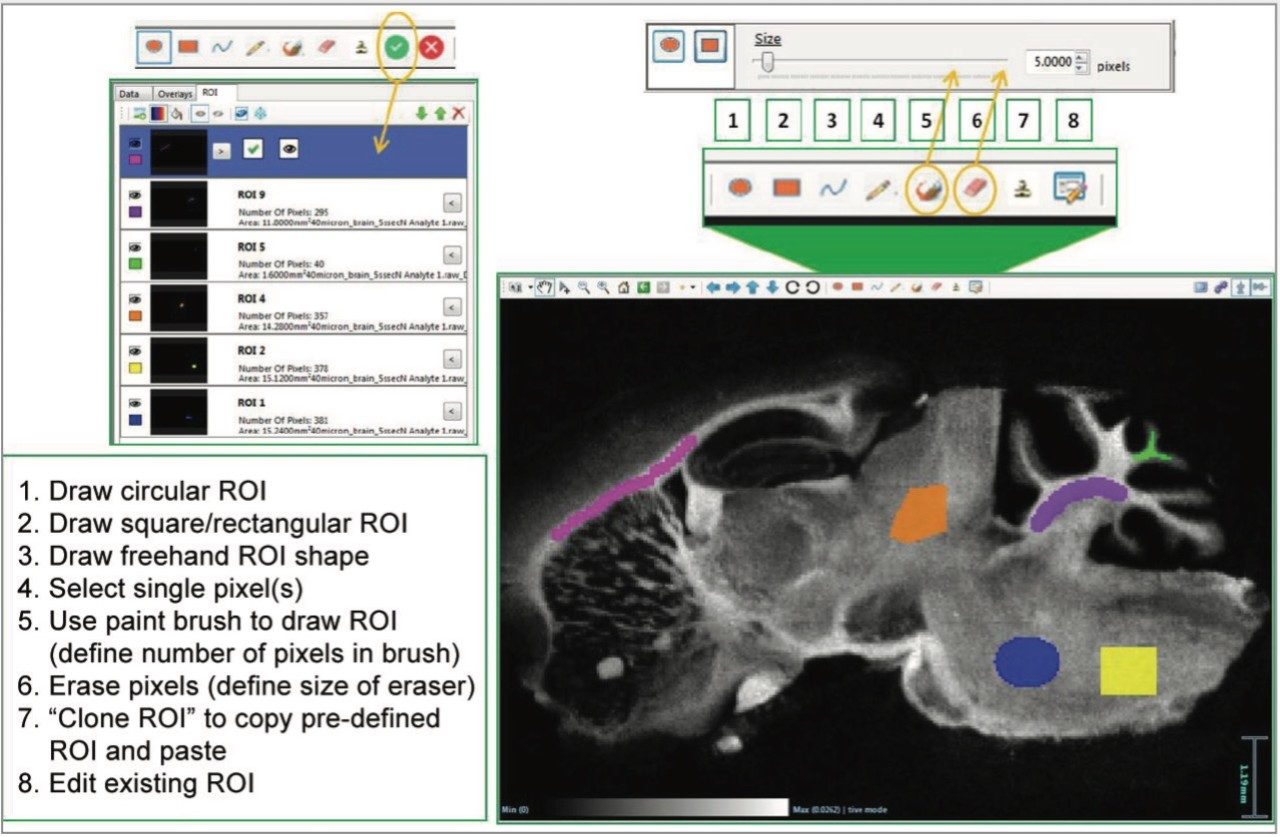
As the ROI is drawn, the number of pixels within the region is tracked. Once the selection of the region has been completed and accepted by the user, it is added in a new tab with its dimensions – which allows for easy management and manipulation of the ROI for analysis.

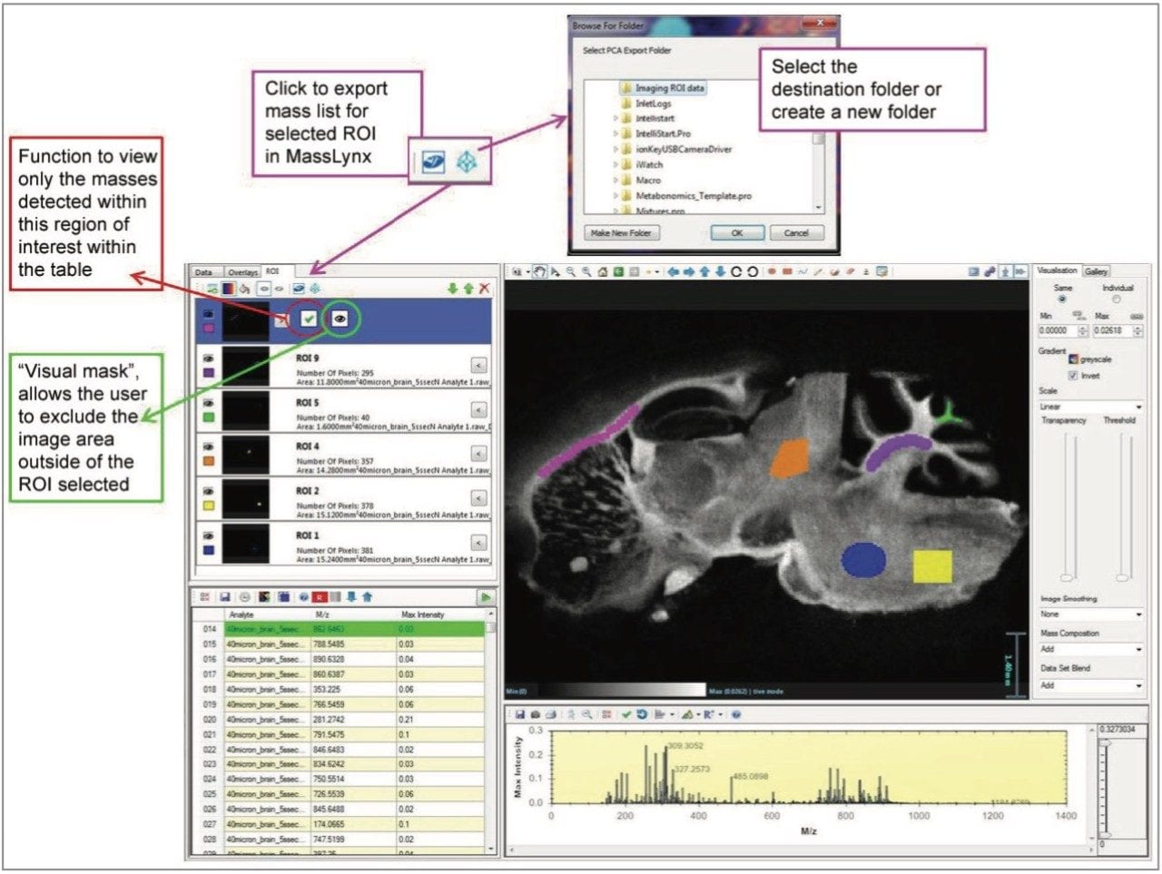
Once the ROIs have been defined, the user has a number of options for further analysis. Both the peak intensities in the analyte table and the MS spectrum can be updated to represent data specific for a given ROI. This is called applying a data mask. Additionally, the spectra within an ROI can be exported and opened in MassLynx MS Software as a combined continuum spectrum, which aids the user in observing any changes in detection of ions across ROI.

Multiple regions from a single image can be selected and the data exported as a spreadsheet format (.csv file) for third-party multivariate analysis software platforms to carry out further statistical analysis. In this case, the intensities for each species within the processed MS imaging dataset (combined ROIs) are averaged, and total ion counts are normalized. ROI can also be exported from multiple sample datasets into a single spreadsheet for multivariate analysis. This easy export of imaging data has been designed for users to take their results and interrogate them externally, increasing flexibility and enhancing the information gained from all experiments.

As a final addition to the ROI functionality, the option for creating a “visual mask” (Figure 2) has been included. This reduces visual complexity by hiding the area outside of a selected ROI from view, allowing examination of discreet regions within an ion image. For consistency, the mass list can also be filtered to display only the m/z values present in a selected ROI.

Figure 2. Workflow for the selection of an ROI, for filtering of the mass list or application of a visual mask. Export processing workflow is also demonstrated.

Conclusion

  • Dedicated ROI tab for easy management of multiple ROI
  • Increased functionality for precise definition when drawing of ROI, even down to a single pixel
  • ROI-masking for visual clarity of the ion images to more easily view specific exclusive regions
  • Export selected ROI back into MassLynx or as a .csv file for further analysis

720005799, September 2016

Zurück zum Seitenanfang Zurück zum Seitenanfang